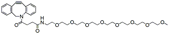
m-PEG12-DBCO is a long chain click chemistry PEG reagents. The DBCO group can react with azides via a copper-free Click Chemistry reaction to form a stable triazole. This reagent can be applied to situations when copper is a concern for DBCO-activated PEGylation. Reagent grade, for research purpose. Please contact us for GMP-grade inquiries.
Detail
m-PEG12-DBCO is a long chain click chemistry PEG reagents. The DBCO group can react with azides via a copper-free Click Chemistry reaction to form a stable triazole. This reagent can be applied to situations when copper is a concern for DBCO-activated PEGylation. Reagent grade, for research purpose. Please contact us for GMP-grade inquiries.
Other Products
R6672C MagPure Pathogen DNA/RNA Enrich Kit (tNGS)
Product Info
Document
Product Info
Introduction
This kit is suitable for extracting total pathogen nucleic acid from a variety of clinical samples such as blood, serum, plasma, swab soaking solution, fluid accumulation and homogenate solution. This kit is designed to remove host cells background nucleic acid and enrich pathogen nucleic acid (including viral/bacterial/fungal DNA/RNA) from the sample. Purified DNA/RNA is ready for downstream applications such as PCR, virus detection, tNGS and other related experiments.
Details
Specifications
Features
Specifications
Main Functions
Extract Pathogen RNA/DNA from 0.5-1.5ml whole blood, plasma, serum, body fluid, homogenate suspension, culture solution, cell suspension, soaking solution or concentrate pathogen solution for tNGS application, remove host background nucleic acid.
Applications
Real Time PCR, biochip analysis, NGS
Products
Pathogen DNA / RNA
Purification method
Polydisperse magnetic beads
Purification technology
Magnetic beads technology
Process method
Manual or automatic
Sample type
whole blood, plasma, serum, body fluid, homogenate suspension, culture solution, cell suspension, soaking solution or concentrate pathogen solution
Sample amount
0.5 – 1.5 ml
Adaptive instrument
Nucleic acid extractor, pipetting workstation
Principle
This product is based on the purification method of high binding magnetic particles. The sample is lysed and digested under the action of lysate and Protease. After adding magnetic particles and binding solution, DNA/RNA will be adsorbed on the surface of magnetic particles, and impurities such as proteins will be removed without adsorption. The adsorbed particles were washed with washing solution to remove proteins and impurities, washed with ethanol to remove salts, and finally DNA/RNA was eluted by Buffer NFW.
Kit Contents
Contents
R667200C
R667202C
Purification Times
24 Preps
96 Preps
2ml Bead Tube (0.4g)
24
96
DNase I (Powder)
10 mg
15 mg
DNase Buffer
5 ml
20 ml
Protease Dissolve Buffer
3 ml
8 ml
Lysis Buffer LBX1
40 ml
180 ml
Buffer TL
5 ml
20 ml
Proteinase K
24 mg
120 mg
MagBind Particles N9
1.2 ml
5 ml
Buffer MLB
30 ml
120 ml
Buffer MW1*
13 ml
110 ml
Buffer MW2*
10 ml
50 ml
Buffer AVE
10 ml
20 ml
Storage and Stability
Proteinase K, DNase I powder and MagPure Particles N9 should be stored at 2–8°C upon arrival. However, short-term storage (up to 8 weeks) at room temperature (15–25°C) does not affect their performance. The remaining kit components can be stored at room temperature (15–25°C) and are stable for at least 18 months under these conditions.
Document
This kit is suitable for extracting total pathogen nucleic acid from a variety of clinical samples such as blood, serum, plasma, swab soaking solution, fluid accumulation and homogenate solution. This kit is designed to remove host cells background nucleic acid and enrich pathogen nucleic acid (including viral/bacterial/fungal DNA/RNA) from the sample. Purified DNA/RNA is ready for downstream applications such as PCR, virus detection, tNGS and other related experiments.
All shipping requirements in one convenient mailer
Leak-proof biohazard bags with adsorbent pads for enhanced safety
Matches the standards of OSHA as well as the IATA regulations
Norgen’s Shipping Accessories provides a safe and convenient tool for shipping non-infectious biological specimens. The accessories are contained in individual SHIP TO envelopes that are used for shipping of both the accessories as well as the individual sample collection device to the donor/user.
Each SHIP TO envelope contains:
A Biohazard Specimen Bag with absorbent pad;
A Bubble Envelope labeled as “Exempt Human Specimen”;
2 blank labels;
Shipping Instructions
The Biohazard Specimen Bag is a sure-seal zipper bag to hold the collected specimen and protect against accidental spills. The bag contains an absorbent pad that can adsorb up to 4 mL of liquid and meets current labeling requirements of OSHA standard 29CFR1910.1030 regarding biohazard symbols. The Bubble Envelope is used as a RETURN envelope for the donor/user to ship the collected sample back to the lab for analysis. The user will first place the collected sample into the Biohazard Specimen Bag, and then place the Biohazard Specimen Bag into the Bubble Envelope. The Bubble Envelope protects against moisture and punctures, and facilitates smooth insertion with convenient self-sealing closure. The 2 labels are used for both the outer SHIP TO envelope and the RETURN bubble envelope. The SHIP TO label will be filled out to contain the mailing information of the donor and the RETURN label will be filled out to contain the address to which the sample should be returned. The labels can be supplied as sheets together with the word template file for ease of printing. Norgen’s Shipping Accessories match the regulations of the International Air Transport Association (IATA).
Champion™ Competent Cells are chemically competent cells, which were prepared by SMOBIO to make E. coli perform excellent transformation efficiency. Standard transformation protocol is recommended for large plasmids or non-ampicillin selection. Time-saving transformation protocol is recommended for simple and rapid transformation. Champion™ Competent Cells are one of the fastest and simplest ready-to-use competent cell products in the world.
Kit contents
Champion™ Competent Cells
pUC19 Control Plasmid (5 μl, 10-4 μg/μl)
Champion™ Transformation Protocol Card
Shipping condition
Throughout the shipping process, the temperature is maintained under -70°C.
Storage and expiration
Champion™ Competent Cells must be stored between -70°C to -80°C. Subsequent freeze-thaw cycles will reduce transformation efficiency. If high efficiency is required for the experiment, do not use aliquots that have gone through several freeze-thaw cycles. The efficiency of Champion™ Competent Cells lasts for 1 year with proper storage.
Document
Champion™ Competent Cells are chemically competent cells, which were prepared by SMOBIO to make E. coli perform excellent transformation efficiency. Standard transformation protocol is recommended for large plasmids or non-ampicillin selection. Time-saving transformation protocol is recommended for simple and rapid transformation. Champion™ Competent Cells are one of the fastest and simplest ready-to-use competent cell products in the world.