

K-INOSL
SKU: 700004304
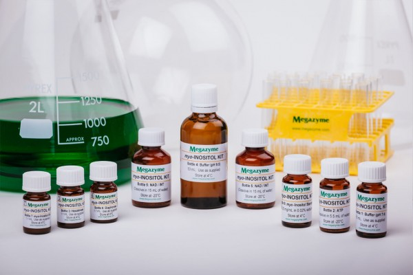
50 assays per kit
| Content: | 50 assays per kit |
| Shipping Temperature: | Ambient |
| Storage Temperature: | Short term stability: 2-8oC, Long term stability: See individual component labels |
| Stability: | > 2 years under recommended storage conditions |
| Analyte: | myo-Inositol |
| Assay Format: | Spectrophotometer |
| Detection Method: | Absorbance |
| Wavelength (nm): | 492 |
| Signal Response: | Increase |
| Linear Range: | 2 to 35 µg of myo-inositol per assay |
| Limit of Detection: | 0.8 mg/L |
| Reaction Time (min): | ~ 30 min |
| Application examples: | Animal feeds, food and other materials. |
| Method recognition: | Novel method |
The myo-Inositol Assay Kit is a reliable and accurate enzymatic UV-method for the specific measurement and analysis of myo-inositol in animal feeds, foods and various other materials.
Phytic acid content of samples with very low levels of free myo-inositol can also be determined using K-INOSL. This can be achieved by measuring the amount of myo-inositol released after de-phosphorylation of phytic acid with the enzymes phytase and alkaline phosphatase, as used with the Megazyme Phytic Acid/Total Phosphorus Assay Kit (K-PHYT).
Not suitable for the determination of myo-inositol in baby formula.
Note for Content: The number of manual tests per kit can be doubled if all volumes are halved. This can be readily accommodated using the MegaQuantTM Wave Spectrophotometer (D-MQWAVE).
Find out more of our alcohol test kits.

Advantages
- Very cost effective
- All reagents stable for > 2 years after preparation
- Only enzymatic kit available
- Rapid reaction
- Mega-Calc™ software tool is available from our website for hassle-free raw data processing
- Standard included


