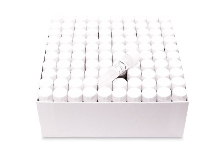
Norgen’s Nuclease-Free Water is free of all DNases, RNases and nucleic acids and is suitable for all molecular biology applications requiring nuclease-free water including PCR, RT-PCR and real-time PCR. Our water is prepared without the use of DEPC, and is ready to use without further treatment.
Applications For use in any molecular biology application
Storage Conditions Norgen’s Nuclease-Free Water should be stored at room temperature. Storage at +4°C or -20°C is optional.
Precautions and Disclaimers This product is designed for research purposes only. It is not intended for human or diagnostic use.
Other Products
Influenza A (H1N1) TaqMan RT-PCR Detection Kits
Product Info
Document
Product Info
Overview
Detection kits for H1N1
CE-IVD marked version available for in vitro diagnostic use
Available in TaqMan format for analysis
Influenza is caused by three immunologic types of RNA viruses (A, B and C) within the Orthomyxoviridae family. Seasonal influenza is typically caused by three major subtypes of hemaglutinin (H1, H2 and H3) and two subtypes of neuraminidase (N1 and N2). A novel sub-type of influenza A virus called pandemic H1N1 2009 virus was identified in Mexico and reported by the CDC and WHO in April, 2009 (Novel swine-origin influenza A (H1N1) virus investigation team, 2009; CDC, 2009; and Fraser et al., 2009). H1N1 2009 is a novel sub-type virus that transmits easily between humans with 21 countries reporting cases within a month of initial identification (CDC, 2009-b). It is essential that public health laboratories around the world undertake detailed surveillance to monitor the spread and impact of pandemic H1N1 2009 virus as well as try to predict future changes in virulence (Fraser et al., 2009). Methods for the rapid diagnosis, case identification and tracking of this novel pathogen in the human population are therefore required to develop appropriate management strategies to mitigate morbidity and mortality.
H1N1 TaqMan RT-PCR Kit, 100 reactions
Ready to use format, including Master Mix for the target and PCR control to monitor for PCR inhibition and validate the quality
Specific Primer and Probe mix for the pathogen/virus/viroid of interest
Primer and Probe mix
Positive and negative control to confirm the integrity of the kit reagents
H1N1 TaqMan RT-PCR Probe/Primer Set and Controls, 100 reactions
Specific Primer/Probe mix and Positive Control for the pathogen/virus/viroid of interest
Nuclease-free water
Can be used together with Norgen’s RT-PCR Master Mix (#28113) or customer supplied master mix
Storage Conditions and Product Stability All kit components can be stored for 1 year after the date of production without showing any reduction in performance.
All kit components should be stored at -20°C upon arrival. Repeated thawing and freezing (> 2 x) of the Master Mix and Positive Control should be avoided, as this may affect the performance of the assay. If the reagents are to be used only intermittently, they should be frozen in aliquots.
The kit offers the unique feature to isolate total RNA including small RNA and DNA from serum and plasma without the need to resort to the cumbersome phenol/chloroform extraction or a time consuming proteinase digest. RNA purified using the kit is ready for applications such as RT-PCR, Northern blotting, poly A+ RNA (mRNA) purification, nuclease protection, and in vitro translation.
Details
Specifications
Features
Specifications
Main Functions
Isolation total RNA and miRNA from 0.2-0.6ml serum and plasma
Applications
Purification method
Mini spin column
Purification technology
Silica technology
Process method
Manual (centrifugation or vacuum)
Sample type
Sample amount
Yield
Elution volume
Time per run
Liquid carrying volume per column
Binding yield of column
Principle
HiPure RNA technology simplifies total RNA isolation. The sample material is denatured in Lysis Buffer. The protein is then precipitated by Protein Precipitation Solution and pelleted by centrifugation. After the removal of protein the binding conditions for nucleic acids are adjusted by adding isopropanol. Total nucleic acids are bound to the column. Optionally, DNA can be removed by an on-column DNase digest. The remaining nucleic acids are washed and eluted with minimal amounts of RNase-Free water.
Kit Contents
Contents
R431402
R431403
Purification Times
50 Preps
250 Preps
HiPure RNA Mini Columns
100
2 x 250
2ml Collection Tubes
100
2 x 250
Buffer CFL
6 ml
30 ml
Buffer CFP
1.8 ml
10 ml
Buffer RWC
20 ml
80 ml
Buffer RW2*
20 ml
2 x 50 ml
RNase Free Water
10 ml
60 ml
Storage and Stability
The kit components can be stored at room temperature (15-25°C) and are stable for at least 18 months under these conditions.
Document
The kit offers the unique feature to isolate total RNA including small RNA and DNA from serum and plasma without the need to resort to the cumbersome phenol/chloroform extraction or a time consuming proteinase digest. RNA purified using the kit is ready for applications such as RT-PCR, Northern blotting, poly A+ RNA (mRNA) purification, nuclease protection, and in vitro translation.
Apoptosis is an essentially normal physiological process that removes now redundant, cells, particularly during embryonic development and early growth. In adult animals the process removes cells that are irreparable. The apoptotic process is also involved in many major diseases such as cancer, where transformed tumour cells have their apoptotic process disabled, permitting cell cycling to continue unchecked. In contrast some forms of senile dementia may result from excessive apoptotic induction of neural cells.
The apoptotic process in mammalian cells is a rapid event (2‐4 hours). Within this short time span an apparently viable cell can be quietly dismantled, to disappear leaving no visible trace of its former existence.
How is apoptosis detected or measured?
An apoptosis cascade of activators, effectors and regulators has been identified. This in turn led to a range of apoptosis assays being devised to detect and monitor these events. Some laboratories will employ two distinct assays, one selected to detect early (initiation) apoptotic events, while a second assay will target a later (execution) event. Apoptosis assays, based on methodology, can be classified into four major inter‐linked groups:
[1] DNA fragmentation (electrophoresis and nick end labelling, TUNEL).
[2] Apoptotic proteases (fluorescently labelled antibodies to the caspases).
[3] Flow cytometric analysis (FACS, incorporating other group assays).
Biocolor’s APOPercentage assay is based on the latter. Further information can be found under the ‘Mode of Action’ Tab.
How does APOPercentage detect apoptosis?
The mammalian cell membrane has been described as a semi‐fluid mosaic structure, composed of phospholipids with a diverse group of inserted proteins and some cholesterol. The phospholipids are the major components of the membrane and are arranged in the form of a ‘bi‐layer’; which is asymmetric in composition, structure, and function.
To ensure normal transmembrane functions the phospholipids must be maintained in an asymmetric composition. The process is regulated by ‘flippases’, which catalyse the active transport of aminophospholipids from the outer to inner monolayer. However, in cells undergoing apoptosis, flippase is overwhelmed by the action of another enzyme, termed ‘floppase’ or ‘scramblase’. The net effect is a scrambling of the phospholipid distribution between the inner and outer monolayers.
Cell membrane changes during apoptosis
The APOPercentage assay utilises an intense, pink-coloured dye reagent which is taken up during in-vitro culture by apoptosis-committed cells. This uptake occurs at the stage of Phosphatidylserine transmembrane movement, as produced by the flipflop mechanism. Dye uptake continues until blebbing occurs. No further dye can then enter the now defunct cell and the dye that has accumulated within the cell is not released (unlike necrotic cells which release dye).
Since the dye reagent is excluded or not retained by healthy or necrotic cells it therefore acts as a specific label for apoptotic cells.
How are APOPercentage-labelled cells quantified?
Labelled apoptosis cells may then by conveniently analysed by the following methods:
Direct Analysis The intense pink colour of the labelled cells can be visually assessed using brightfield microscopy. Apoptosis in substrate-adherent cell populations is therefore readily quantified using image analysis techniques. This technique is the most sensitive with the ability of detecting one single apoptotic cell per well.
Colorimetry protocol Dye that accumulates within apoptotic cells is released into solution via addition of Dye Release Reagent. The concentration of this intracellular dye is then measured at 550nm using a microplate colorimeter/spectrophotometer.
NB: The APOPercentage assay kit does NOT require the use of a Flow Cytometer.
Limit of Detection
A single cell (via image analysis method)
Detection Method
Colorimetric (550nm) (Endpoint) or Image Analysis based
Measurements per kit
Sufficient for 4×24 well plates or 6×96 well plates
Suitable Samples
Adherent mammalian cells (in-vitro)
APOPercentage kit contents:
1. APOPercentage Dye (1x5ml)
2. Dye Release Reagent (1x150ml)
3. Phosphate Buffered Saline (PBS) (1x120ml)
4. 24-well starter plate.
5. Assay kit manual.
The Colorimetric Protocol requires a Microplate Colorimeter / Spectrophotometer.
Additional 96-well plates will be required for use when reading dye absorbance values.
The Direct Detection Protocol Requires an inverted stage microscope with an attached digital camera.
NB: Additional reagents (typically culture medium and suitable apoptosis treatments) may be required for sample preparation prior to assay. Consult manual or contact us for further details.
Document
The APOPercentage™ Apoptosis kit is a dye-based, colorimetric assay for detection and measurement of apoptosis (programmed cell death) during in-vitro cell culture.