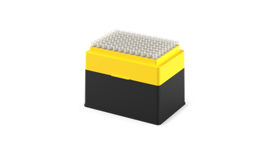
200 µL filtered pipette tips for the Opentrons Flex, provided sterile in racks or blister packs (refills). Tips are clear, non-conducting polypropylene tips, free of human DNA, PCR inhibitors, RNase/DNase, pyrogens, and endotoxins. Opentrons Flex Tips and Racks are autoclavable.
Detail
200 µL filtered pipette tips for the Opentrons Flex, provided sterile in racks or blister packs (refills). Tips are clear, non-conducting polypropylene tips, free of human DNA, PCR inhibitors, RNase/DNase, pyrogens, and endotoxins. Opentrons Flex Tips and Racks are autoclavable.
Other Products
Escherichia coli (E.coli) O157:H7 Quantified Bacterial DNA Standards
Product Info
Document
Product Info
Overview
Quantified standard to be used as a positive control or PCR quantification standard
Vigorously quantified using multiple methods
Includes partial sequence of STX-1 subunit A, and STX-2 subunit A genes (please refer to product insert for more details)
Compatible with E. coli O157:H7 TaqMan PCR Detection Kit and E. coli O157:H7 TaqMan PCR Detection Probe/Primer and Control Set
E. coli O157:H7 is a rod-shaped, gram negative bacterium. It is an enterohemorrhagic strain of the common E. coli bacterium and infection by the O157:H7 strain is commonly associated with hemorrhagic colitis. E. coli O157:H7 is recognized by its somatic (cell wall) antigen (O157) and its flagella antigen (H7). In addition, E. coli O157:H7 is known to produce Shiga-like toxins, which cause severe symptoms. While most patients can recover from the infection, up to 15% of the patients may develop hemolytic uremic syndrome, a type of kidney failure that could be fatal. Infection of E. coli O157:H7 usually results from consumption of poorly prepared food including undercooked meat (particularly ground beef), untreated water or raw unpasteurized milk.
Norgen’s E. coli O157:H7 Quantified Bacterial DNA Standards are cloned fragments of the two Shiga-like toxin regions purified using Norgen’s sample preparation technology. They include partial sequences of STX-1 subunit A gene and STX-2 subunit A gene. Please refer to Appendix A for sequence information. The purified DNA is quantified vigorously using multiple methods including spectrophotometry, gel densitometry and real-time PCR. It is intended to be used as positive controls or PCR quantification standards for E. coli O157:H7. This product is compatible with Norgen Biotek’s E. coli O157:H7 Taqman PCR Detection Kit (Cat. TM41350) and E. coli O157:H7 Taqman PCR Detection Probe/Primer and Control Set (Cat. TM41310).
Storage Conditions Upon receipt, store Norgen’s E. coli O157:H7 Quantified Bacterial DNA Standards at -20°C or lower. Avoid multiple freeze-thaw cycles. If needed, prepare smaller working aliquots and store at -20°C or lower.
[PM5200] ExcelBand™ 3-color Pre-Stained Protein Ladder, Broad Range (3.5-245 kDa), 250 μl x 2
Product Info
Document
Product Info
Description
The PM5200 3-color Pre-Stained Protein Ladder Broad Range is a ready-to-use three-color protein standard with 15 pre-stained proteins covering a wide range of molecular weights from 5 to 245 kDa in Tris-Glycine Buffer (3.5 to 235 kDa in Bis-Tris (MOPS) buffer and Bis-Tris (MES) buffer). Proteins are covalently coupled with different chromophores for easy identification of bands, with three reference proteins carrying enhanced intensity corresponding to a blue band at 20 kDa, green at 40 kDa, and red at 75 kDa, respectively, as separated on SDS-PAGE (Tris-Glycine buffer). The PM5200 3-color Pre-Stained Protein Ladder Broad Range is designed for monitoring protein separation during SDS-polyacrylamide gel electrophoresis, verification of Western transfer efficiency on membranes (PVDF, nylon, or nitrocellulose) and for approximating the size of proteins.
Features
Ready-to-use — Premixed with a loading buffer for direct loading, no need to boil.
Three reference bands — 75 kDa (red), 40 kDa (green), and 20 kDa (blue)
Contents
Approximately 0.1~0.4 mg/ml of each protein in the buffer (20 mM Tris-phosphate (pH 7.5 at 25°C), 2% SDS, 0.2 mM DTT, 3.6 M urea, and 15% (v/v) glycerol).
Quality Control
Under suggested conditions, PM5200 ExcelBand™ 3-color Pre-Stained Protein Ladder Broad Range resolves 15 major bands in SDS-PAGE (Tris-Glycine, MOPS, and MES buffer) and after Western blotting to nitrocellulose membrane.
Storage
4°C for 3 months -20°C for long term storage
Document
The PM5200 3-color Pre-Stained Protein Ladder Broad Range is a ready-to-use three-color protein standard with 15 pre-stained proteins covering a wide range of molecular weights from 5 to 245 kDa in Tris-Glycine Buffer (3.5 to 235 kDa in Bis-Tris (MOPS) buffer and Bis-Tris (MES) buffer). Proteins are covalently coupled with different chromophores for easy identification of bands, with three reference proteins carrying enhanced intensity corresponding to a blue band at 20 kDa, green at 40 kDa, and red at 75 kDa, respectively, as separated on SDS-PAGE (Tris-Glycine buffer). The PM5200 3-color Pre-Stained Protein Ladder Broad Range is designed for monitoring protein separation during SDS-polyacrylamide gel electrophoresis, verification of Western transfer efficiency on membranes (PVDF, nylon, or nitrocellulose) and for approximating the size of proteins.
Introducing the Fastin Assay Kit: Your Straightforward Solution for Elastin Quantification! Our user-friendly kit utilizes a dye-based method to measure elastin from in-vivo and in-vitro sources. It can be used to quantify various elastin forms, spanning from immature tropoelastin to mature, ‘insoluble’ elastin fibers.
Colorimetric Detection (513nm) (Endpoint)
Understanding Elastin: The Key to Tissue Flexibility
Tissues like lungs and arteries must maintain the ability to stretch and recoil repeatedly throughout an organism’s life. Elastin, a mature protein, is responsible for this elasticity and is usually present as insoluble fibers within the ECM. During development, these fibers are initially formed from a soluble precursor called tropoelastin.
What is the Fastin Assay?
The Biocolor Fastin assay is a user-friendly, dye-based means of quantifying elastins derived from both in-vivo and in-vitro sources. A variety of elastin forms can be assayed, from immature tropoelastin to mature ‘insoluble’ elastin fibres.
Further information on how the assay works can be found on the ‘Mode of Action‘ tab.
A list of suggested sample types can be found under the ‘Assay Specification‘ tab.
How does the Fastin assay detect Elastin?
The Fastin Dye Reagent contains an elastin-binding synthetic porphyrin, TPPS (5,10,15,20-tetraphenyl- 21H,23H-porphine). This affinity of TPPS for elastin was first observed when used as a ‘vital stain’ on live animals. Most tissues took up the dye initially but only elastin retained the TPPS molecules over time. [Winkelman, J. (1962), Cancer Res. 22, 589-596; Winkelman, J & Spicer, S. (1962), Stain Technol. 37, 303-305].
It has been proposed that the elastin binding of TPPS may be due to the retention of the acidic dye (which contains four charged sulfate groups) by the basic amino acid side chain residues of elastin.
How does the Fastin assay work?
Step 1. Incubation of samples containing soluble elastin with the Fastin Dye Reagent causes an elastin-dye complex to form. This insoluble complex then precipiates.
Step 2. Dye-labelled elastin is then isolated by centrifugation and the unbound dye removed. Elastin-bound dye is then eluted and measured spectrophotometrically.
Step 3. The elastin content of unknown samples can be calculated by comparison against a calibration curve prepared using a standard comprising water-soluble elastin (supplied with the kit).
Assay range
50 – 500µg/ml
Limit of Detection
50µg/ml
Detection Method
Colorimetric Detection (513nm) (Endpoint)
Measurements per kit
110 in total (allows a maximum of 48 samples to be run in duplicate alongside a standard curve).
Suitable Samples
In-vivo: tissues and fluids. Insoluble elastin will first require conversion to water soluble α-elastin using the oxalic acid reagents and extraction protocol supplied with the kit.
In-vitro: Elastin produced by cells during 2D/3D cell culture. NB elastin in conditioned cell media is typically below the detection limit of the kit.
Precautions
This kit is designed for research use only. Not for use in diagnostic procedures. Kit requires access to a centrifuge, heated water bath or block, as well as a spectrophotometer or colorimeter capable of absorbance detection at 513nm. Specific sample preparation protocols may require customer to provide further reagents, consult assay manual for further information.
Fastin elastin kit contents:
1. Dye Reagent (1x110ml)
2. α-elastin Reference Standard (1x5ml, 1.0 mg/ml soluble Bovine elastin)
6. 1.5ml micro-centrifuge tubes for dye-labelling reaction.
7. Assay kit manual
NB: Additional reagents may be required for sample preparation prior to assay. Consult manual or contact us for further details.
Document
Introducing the Fastin Assay Kit: Your Straightforward Solution for Elastin Quantification! Our user-friendly kit utilizes a dye-based method to measure elastin from in-vivo and in-vitro sources. It can be used to quantify various elastin forms, spanning from immature tropoelastin to mature, ‘insoluble’ elastin fibers. Colorimetric Detection (513nm) (Endpoint)