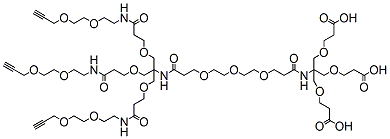
PEG3-(Amino-Tri-(Propargyl-PEG2-ethoxymethyl)-methane)-(Amino-Tri-(carboxyethoxymethyl)-methane) is reactive with azide-bearing compounds or biomolecules via copper catalyzed azide-alkyne Click Chemistry to yield a stable triazole linkage. The terminal carboxylic acid groups can react with primary amino groups in the presence of activators (e.g. HATU) to form a stable amide bond.
Detail
PEG3-(Amino-Tri-(Propargyl-PEG2-ethoxymethyl)-methane)-(Amino-Tri-(carboxyethoxymethyl)-methane) is reactive with azide-bearing compounds or biomolecules via copper catalyzed azide-alkyne Click Chemistry to yield a stable triazole linkage. The terminal carboxylic acid groups can react with primary amino groups in the presence of activators (e.g. HATU) to form a stable amide bond.
Other Products
R6641 MagPure Plant RNA Kit
Product Info
Document
Product Info
Introduction
This product supplies a simple and rapid extraction of total RNA from plant and fungal samples. The kit is based on super paramagnetic particles purification technology, no phenol-chloroform extraction or alcohol precipitation. The whole extraction process takes only 60 minutes. Purified RNA is ready for downstream applications such as RT-PCR, virus RNA testing and so on. MagPure RNA Kits buffers can be used for both manual extraction process and automatic nucleic acid extraction machines. This Kits is suitable for extracting RNA from ≤5×10⁶ cultured cells, 20mg tissue and <50mg plant samples.
Details
Specifications
Features
Specifications
Main Functions
Isolation total RNA from 50mg plant using magnetic particles
Applications
RT-PCR, cDNA synthesis, second generation sequencing
Purification method
Polydisperse magnetic beads
Purification technology
Magnetic beads technology
Process method
Manual or automatic
Sample type
Plant and fungus samples
Sample amount
≤50mg
Yield
2-100μg
Time per run
≤60 minutes
Principle
This product is based on the purification method of high binding magnetic particles. The sample is lysed and digested under the action of lysate and Protease. After adding magnetic particles and binding solution, RNA will be adsorbed on the surface of magnetic particles, and impurities such as proteins will be removed without adsorption. The adsorbed particles were washed with washing solution to remove proteins and impurities, washed with ethanol to remove salts, and finally RNA was eluted by Elution Buffer.
Advantages
High quality – high purity total RNA can be directly used in various sensitive downstream applications
Safe – no phenol chloroform extraction required
Fast – several samples can be extracted in 60 minutes by column method
Universal – two lysates suitable for most plant or fungal tissue samples
Kit Contents
Contents
R664101
R664102
R664103
Purification Times
48 Preps
96 Preps
480 Preps
MagPure RNA Particles
1.7 ml
3.5 ml
18 ml
DNase I
600 μl
2 x 600 μl
10 x 600 μl
DNase Buffer
30 ml
40 ml
200 ml
Buffer PRC1
40 ml
70 ml
350 ml
Buffer RL
40 ml
70 ml
350 ml
Buffer MCB*
18 ml
30 ml
150 ml
Buffer MW1*
22 ml
44 ml
220 ml
Buffer MW2*
20 ml
50 ml
2 x 100 ml
RNase Free Water
10 ml
15 ml
120 ml
Storage and Stability
MagPure RNA Particles should be stored at 2–8°C upon arrival. DNase I should be stored at -20°C. However, short-term storage (DNase I up to 1 weeks, MagPure RNA Particles up to 8 weeks) at room temperature (15–25°C) does not affect their performance. The remaining kit components can be stored at room temperature (15–25°C) and are stable for at least 18 months under theseconditions.
Document
This product supplies a simple and rapid extraction of total RNA from plant and fungal samples. The kit is based on super paramagnetic particles purification technology, no phenol-chloroform extraction or alcohol precipitation. The whole extraction process takes only 60 minutes. Purified RNA is ready for downstream applications such as RT-PCR, virus RNA testing and so on. MagPure RNA Kits buffers can be used for both manual extraction process and automatic nucleic acid extraction machines. This Kits is suitable for extracting RNA from ≤5×10⁶ cultured cells, 20mg tissue and
DBCO-PEG23-amine is a PEG linker which contains DBCO and amine moieties. The DBCO group is commonly used in copper-free Click Chemistry reactions. The amine group is reactive with carboxylic acids, activated NHS esters, carbonyls (ketone, aldehyde) etc. The hydrophilic PEG spacer increases the water solubility of the compound. Reagent grade, for research purpose. Please contact us for GMP-grade inquiries.
Document
DBCO-PEG23-amine is a PEG linker which contains DBCO and amine moieties. The DBCO group is commonly used in copper-free Click Chemistry reactions. The amine group is reactive with carboxylic acids, activated NHS esters, carbonyls (ketone, aldehyde) etc. The hydrophilic PEG spacer increases the water solubility of the compound. Reagent grade, for research purpose. Please contact us for GMP-grade inquiries.