[PM2600] ExcelBand™ 3-color High Range Protein Marker (9-245 kDa), 250 μl x 2
Facebook
X
Pinterest
Email
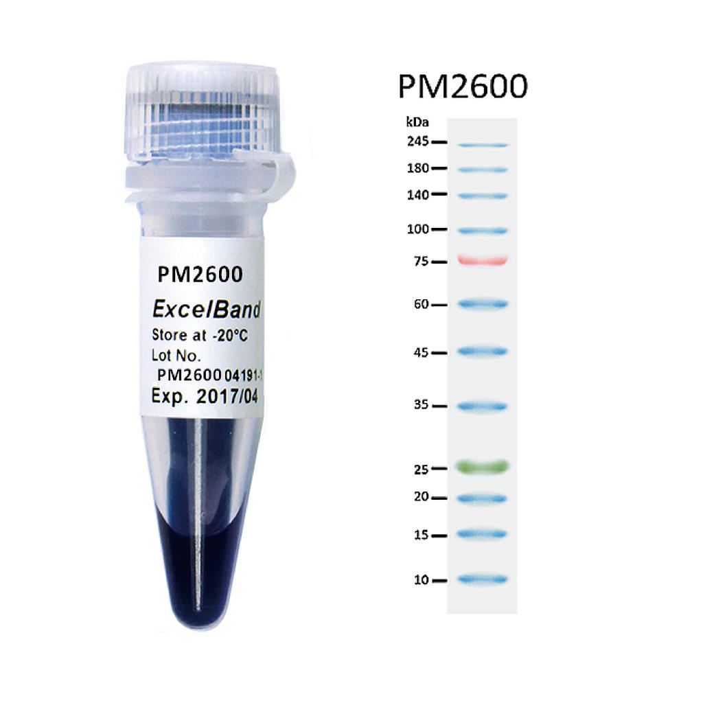
The PM2600 ExcelBand™ 3-color High Range Protein Marker is a ready-to-use three-color protein standard with 12 pre-stained proteins covering a wide range of molecular weights from 10 to 245 kDa in Tris-Glycine Buffer (9 to 235 kDa in Bis-Tris (MOPS) buffer and 10 to 235 kDa in Bis-Tris (MES) buffer). Proteins are covalently coupled with a blue chromophore except for two reference bands (one green and one red band at 25 kDa and 75 kDa respectively) when separated on SDS-PAGE (Tris-Glycine buffer). The PM2600 3-color High Range Protein Marker is designed for monitoring protein separation during SDS-polyacrylamide gel electrophoresis, verification of Western transfer efficiency on membranes (PVDF, nylon, or nitrocellulose) and for approximating the size of proteins.
Detail
Description
The PM2600 ExcelBand™ 3-color High Range Protein Marker is a ready-to-use three-color protein standard with 12 pre-stained proteins covering a wide range of molecular weights from 10 to 245 kDa in Tris-Glycine Buffer (9 to 235 kDa in Bis-Tris (MOPS) buffer and 10 to 235 kDa in Bis-Tris (MES) buffer). Proteins are covalently coupled with a blue chromophore except for two reference bands (one green and one red band at 25 kDa and 75 kDa respectively) when separated on SDS-PAGE (Tris-Glycine buffer). The PM2600 3-color High Range Protein Marker is designed for monitoring protein separation during SDS-polyacrylamide gel electrophoresis, verification of Western transfer efficiency on membranes (PVDF, nylon, or nitrocellulose) and for approximating the size of proteins.
Features
Ready-to-use — Premixed with a loading buffer for direct loading, no need to boil.
Two reference bands — 75 kDa (red) and 25 kDa (green)
Contents
Approximately 0.1~0.4 mg/ml of each protein in the buffer (20 mM Tris-phosphate (pH 7.5 at 25°C), 2% SDS, 0.2 mM DTT, 3.6 M urea, and 15% (v/v) glycerol).
Quality Control
Under suggested conditions, PM2600 ExcelBand™ 3-color High Range Protein Marker resolves 12 major bands in 15% SDS-PAGE (Tris-Glycine buffer, MOPS, and MES buffer) and after Western blotting to nitrocellulose membrane.
Storage
4°C for 3 months -20°C for long term storage
Other Products
D3117 HiPure Tissue&Blood DNA 96 Kit
Product Info
Document
Product Info
Introduction
Blood samples contain rich DNA, including mitochondrial DNA, genomic DNA, circulating DNA (mostly released into blood after tumor cell apoptosis) in white blood cells, as well as parasitic viral or microbial DNA. These DNA are important parameters in clinical testing or diagnosis, which are also valuable materials for medical research. There are three main issues with extracting DNA from blood samples:
1. The sample is highly infectious, posing great harm to operators and the environment.
2. The source of DNA is complex and aportion of the nucleic acid, such as viral DNA or free DNA, may be lost during the operation, leading to downstream detection failure;
3. Blood sample contains a large amount of impurities and inhibitory factors.
Currently there are many methods available for extracting DNA from whole blood samples, such as phenol chloroform extraction, salting out method, etc. However, these methods require pre-treatment of blood sample, which removes red blood cells and isolate white blood cells in the first step. Due to the requirement that it cannot inactivate or kill pathogens during the process of removing red blood cells, the waste liquid (red blood cell lysate) and consumables may be contaminated by pathogens and become infectious, posing a danger to the entire laboratory environment and operators. In addition, during the process of removing red blood cells, useful nucleic acid information such as viruses, microorganisms, or circulating DNA is also lost, leading to experiment or detection failures.
The HiPure Blood DNA Kits series provided by Magen Company uses silica gel column purification technology, which can directly lyse whole blood samples without the need for white blood cell separation. Whole blood samples are directly mixed with lysates and proteases, resulting in the inactivation of pathogens, greatly reducing the infectivity, environmental pollution, and the chance of operators being infected. Due to the direct lysis and digestion of samples, except lymphocyte DNA, other circulating DNA as well as DNA from viruses and microorganisms, can also be recovered.
This product provides fast and easy methods for purification of total DNA for reliable PCR and Southern blotting. Total DNA (e.g., genomic, viral, mitochondrial) can be purified from tissue, whole blood, plasma, serum, buffy coat, bone marrow, other body fluids, lymphocytes, cultured cells.
Details
Specifications
Features
Specifications
Main Functions
Isolation total DNA from blood, tissue, culture cells, swab, blood spots using 96 plate
Applications
PCR, southern bolt and virus detection, etc
Purification method
96 well plate
Purification technology
Silica technology
Process method
Manual (centrifugation or vacuum)
Sample type
Blood, serum, plasma, milk, saliva, and other liquid samples and cultured cells
Sample amount
Elution volume
Time per run
Liquid carrying volume per column
Binding yield of column
Principle
This product is based on silica column purification. The sample is lysed and digested with lysate and protease, DNA is released into the lysate. Transfer to an adsorption column. Nucleic acid is adsorbed on the membrane, while protein is not adsorbed and is removed with filtration. After washing proteins and other impurities, Nucleic acid was finally eluted with low-salt buffer (10mm Tris, pH9.0, 0.5mm EDTA).
Advantages
High quality DNA – meet a variety of downstream applications, including PCR, qPCR, enzyme digestion, hybridization, etc.
High throughput – 96 samples can be processed simultaneously
Kit Contents
Contents
D311701
D311702
Purification Times
1 x 96
4 x 96
HiPure gDNA Plate
1
4
96 well Plate (2.2ml)
1
4
1.6ml Collection Plate
1
4
0.5ml Collection Plate
1
4
Silicon Seal Tape
1
4
Seal Film
5
25
Buffer ATL
30 ml
100 ml
Buffer AL
30 ml
100 ml
Buffer DW1
60 ml
250 ml
Buffer GW2
50 ml
2 x 100 ml
Proteinase K
50 ml
200 ml
Protease Dissolve Buffer
5 ml
15 ml
Buffer AE
30 ml
120 ml
Storage and Stability
Proteinase K should be stored at 2-8°C upon arrival. However, short-term storage (up to 12 weeks) at room temperature (15-25°C) does not affect their performance. The remaining kit components can be stored at room temperature (15-25°C) and are stable for at least 18 months under these conditions.
Blood samples contain rich DNA, including mitochondrial DNA, genomic DNA, circulating DNA (mostly released into blood after tumor cell apoptosis) in white blood cells, as well as parasitic viral or microbial DNA. These DNA are important parameters in clinical testing or diagnosis, which are also valuable materials for medical research. There are three main issues with extracting DNA from blood samples:
E. coli O157:H7 is a rod-shaped, gram negative bacterium. It is an enterohemorrhagic strain of the common E. coli bacterium and infection by the O157:H7 strain is commonly associated with hemorrhagic colitis. E. coli O157:H7 is recognized by its somatic (cell wall) antigen (O157) and its flagella antigen (H7). In addition, E. coli O157:H7 is known to produce Shiga-like toxins, which cause severe symptoms. While most patients can recover from the infection, up to 15% of the patients may develop hemolytic uremic syndrome, a type of kidney failure that could be fatal. Infection of E. coli O157:H7 usually results from consumption of poorly prepared food including undercooked meat (particularly ground beef), untreated water or raw unpasteurized milk.
E.coli O157:H7 TaqMan PCR Kit, 100 reactions
Ready to use format, including Master Mix for the target and PCR control to monitor for PCR inhibition and validate the quality
Specific Primer and Probe mix for the pathogen/virus/viroid of interest
Primer and Probe mix
Positive and negative control to confirm the integrity of the kit reagents
E.coli O157:H7 TaqMan PCR Probe/Primer Set and Controls, 100 reactions
Specific Primer/Probe mix and Positive Control for the pathogen/virus/viroid of interest
Nuclease-free water
Can be used together with Norgen’s PCR Master Mix (#28007) or customer supplied master mix
For research use only and NOT intended for in vitro diagnostics.
Storage Conditions and Product Stability All kit components can be stored for 2 years after the date of production without showing any reduction in performance.
All kit components should be stored at -20°C upon arrival. Repeated thawing and freezing (> 2 x) of the Master Mix and Positive Control should be avoided, as this may affect the performance of the assay. If the reagents are to be used only intermittently, they should be frozen in aliquots.
Heat sealing offers a 100% effective method of plate sealing, for complete seal integrity, as well as being quick and cost effective.
Our Individual Access Peel Heat Seal is a laminate seal compatible with polypropylene plates, featuring 96 individual foil seal spots or 12 strips of individual spots on a removable backing.
These seals result in individually sealed tubes/strips, and they can be removed from polypropylene plates by peeling, even with a plate which has been removed directly from -80°C storage.
Individual Access Peel Heat Seal forms a complete seal to a plate enabling very low temperature uses, including very low temperature storage, and high temperature uses, such as PCR (when used with a pressurized heated lid).
The seal demonstrates moderate solvent resistance and can be utilized for short term compound storage at room temperature.
This seal is available as sheets, for use with manual and semi-automated sealers, such as our Semi-Automated Sheet Heat Sealer (using the 59-2005 Individual Access adapter).
Document
Heat sealing offers a 100% effective method of plate sealing, for complete seal integrity, as well as being quick and cost effective.