ProbeSure Multiplex Master Mix is an enhanced version of ProbeSure Master Mix, formulated to enable users to analyse up to four targets in one reaction well. For example, two bi-allelic SNPs or one reference gene and a further three genes of interest.
Users will require a plate reader capable of reading FAM, HEX, ATTO 550, ATTO 647N and ATTO 633 (the wavelengths of each of these can be found in our ProbeSure Multiplex Master Mix User Guide). ProbeSure Multiplex Master Mix is supplied at 2x concentration for convenience and is supplied with the ATTO 633 normalising dye at either high level (500 nM final concentration), low level (25 nM final concentration) or without ATTO 633.
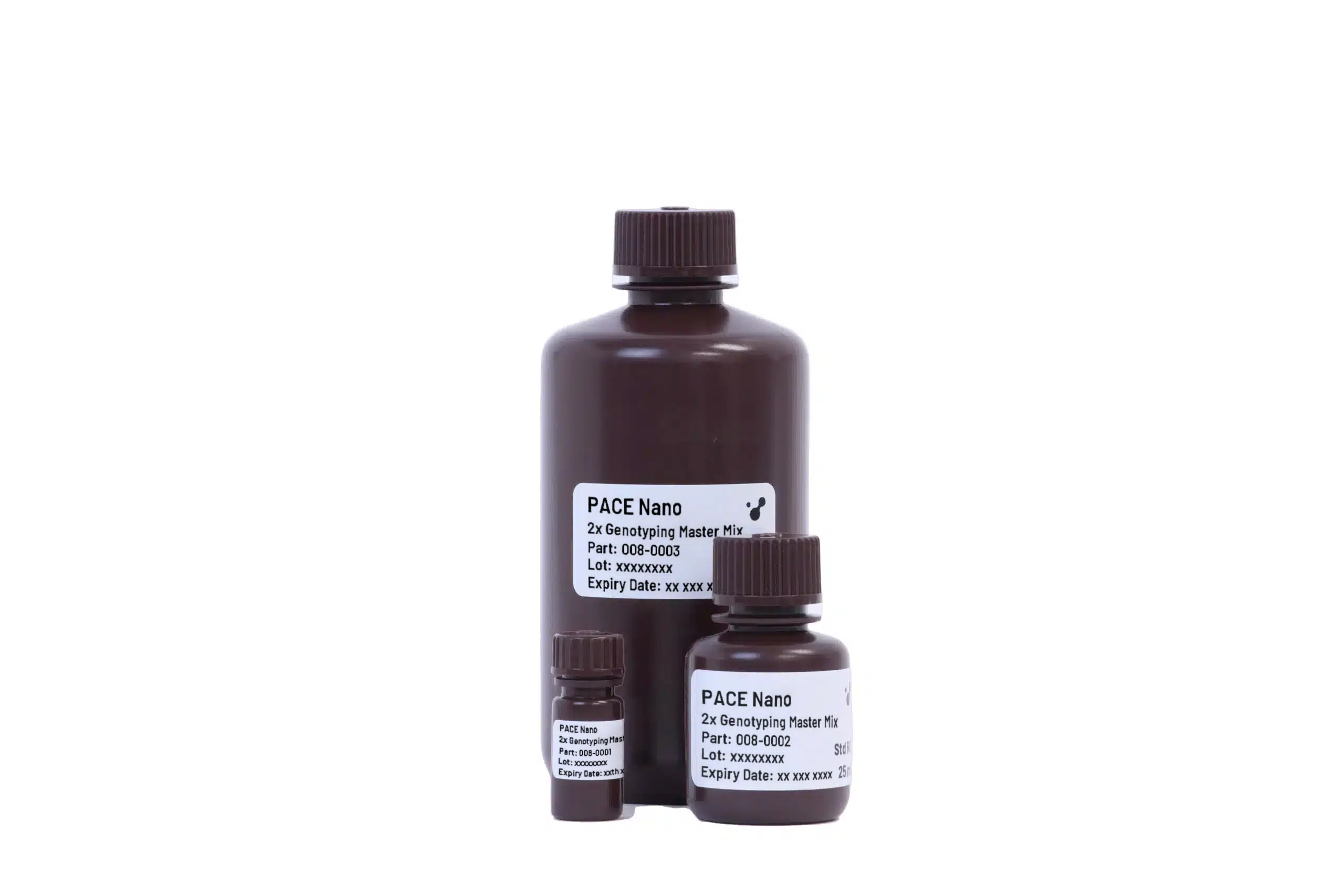
Other Products
Cylindrospermopsin ELISA Kit
Product Info
Document
Product Info
Format: 96-well microtiter plate (12 test strips of 8 wells)
The Cylindrospermopsin plate kit is a competitive enzyme-labeled immunoassay. The Cylindrospermopsin sample extract and calibrators are pipetted into the test wells followed by the Cylindrospermopsin antibody into the test wells to initiate the reaction. During the 30 minutes incubation period, Cylin-drospermopsin from the sample and Cylindrospermopsin antigen compete for binding to the Cylindrosper-mopsin antibody. The Cylindrospermopsin antibody is captured on the walls of the test well. Following this 30-minute incubation, the contents of the wells are removed and the wells are washed to remove any unbound Cylindrospermopsin and free Cylindrospermopsin antibody. After wash, 1X HRP-conjugated Antibody#2 is added for 30 minutes incubation. The wells are washed afterwards, and a clear substrate is then added to the wells and any bound enzyme conjugate causes the conversion to a blue color. Following a 15-minute incubation, the reaction is stopped and the amount of color in each well is read. The color of the unknown samples is compared to the color of the calibrators and the Cylindrospermopsin concentration of the samples is derived.
Format:
• 96-well microtiter plate (12 test strips of 8 wells)
Cat.# 20106S, 20106L: Size range 450-750 bp (ideal for NGS library size selection)
Product Info
Document
Product Info
The series of DNA Size Selection Kits (Magnetic Beads) were developed for DNA size selection using magnetic beads. A total of 11 kits are available, with different selection ranges spanning from 50 bp to over 10 kb. The kits provide a simple and quick approach for the enrichment of a specific range of DNA fragments. The kit workflow allows double-sided or single-sided size selection for specific size cutoffs.
Gel images of different ranges of size selection. Sheared human genomic DNA was used as input.
.
DNA size selection is a selective capture of DNA fragments of a specific range of size for next-generation sequencing (NGS) library preparations, PCR, ChIP assay, DNA ligations, endonuclease digestions, adapter removal, and other genomics and molecular biology applications. DNA size selection is preferred after NGS library prep in most of the cases. The NGS library preparation is related to the quality of the sequencing data. Precise NGS library size selection can increase sequencing efficiency, improve data quality, and reduce costs.
There are two types of sequencing technologies: short-read sequencing and long-read sequencing. Short-read sequencing uses DNA libraries that contain small insert DNA fragments of similar sizes, usually several hundred base pairs. The sequencing efficiency can be improved if the DNA size selection is in the right range. Cat.# 20104S and 20104L are the best kits for NGS library size selection of illumina paired-end 100 (PE100) sequencing with 100-200 bp library inserts; Cat.# 20105S and 20105L are the best kits for NGS library size selection of illumina paired-end 150 (PE150) sequencing with 150-300 bp library inserts; and Cat.# 20106S and 20106L are the best kits for NGS library size selection of illumina paired-end 300 (PE300) sequencing with 300-600 bp library inserts.
Long-read sequencing uses a large DNA fragment as input and makes very long reads. Usually, library size selection is preferred to remove smaller fragments. Cat.# 20110S and 20110L are the best kits for long-read sequencing size selection with DNA sizes >5 kb, and Cat.# 20111S and 20111L are the best kits for long-read sequencing size selection with DNA sizes >10 kb.
The magnetic beads technology uses paramagnetic particles, also known as SPRI (Solid Phase Reversible Immobilization) beads, to bind DNA reversibly and selectively. DNA fragments can be size-selected and purified by changing the properties of the magnetic beads or SPRI beads. The magnetic beads can easily separate the beads-binding DNA from the contaminants and unwanted components in the samples. The samples after DNA size selection are free of contaminants such as buffer components, enzymes, proteins, salts, dNTPs, primers, and adapters. Our proprietary magnetic beads reagents improve yield, selectivity, and reproducibility.
Specific DNA fragments at a certain length range can be purified simply using magnetic separation with different beads components, avoiding tedious and time-consuming gel extraction and column-based purification. The magnetic beads method is popular for common DNA size selection, including library size selection. The first beads-binding step, referred to as the right-side clean-up, removes large DNA fragments. The large DNA fragments are bound to the beads and are discarded. The desired DNA fragments in the supernatant are transferred to a new well, and new beads are added to the supernatant for the second beads-binding, referred to as the left-side clean-up. The double-size selected DNA fragments are eluted after ethanol rinsing.
DNA size selection with dual clean-ups.
.
A single clean-up is needed for DNA size selection with large fragments. In this case, only the large DNA fragments are bound to the beads. The selected larger DNA fragments are eluted after ethanol rinsing.
DNA size selection with single clean-up for >5 kb and >10 kb DNA.
.
Features of DNA size selection and library size selection
High specificity and high recovery of size selection
11 selection ranges are available, including 5 ranges for NGS library size selection
50-100 bp
100-200 bp
200-500 bp
250-350 bp: ideal for illumina PE100 sequencing
300-450 bp: ideal for illumina PE150 sequencing
450-750 bp: ideal for illumina PE300 sequencing
500-1000 bp
1-3 kb
1-5 kb
>5 kb: ideal for long-read sequencing
>10 kb: ideal for long-read sequencing
Fast and simple
20-min protocol
No gel purification required
No columns required
No centrifugation required
Efficient removal of contaminants and unwanted components
Convenient – With the ready-to-use Master Mix, the user needs only to add template to the master mix and enzyme in order to set up the reverse transcriptase reaction
Time Savings – Set up RT reactions in a shorter time since less pipetting steps are required
Cost Efficient – No need to buy separate enzymes, dNTPs and buffers. All are included with the ready-to-use Master Mix kits
High Sensitivity and Yield – the optimized Master Mix allows for highly sensitive amplifications with high yields of PCR products
Robust Enzyme – broad range of working temperatures from 37°C to 60°C. Capable of amplifying difficult templates with a high degree of reproducibility
TruScript Reverse Transcriptase is Available in Three Convenient Formats:
This kit contains 5X RT Buffer and a vial of TruScript Enzyme Mix (200 units/µL). This enzyme can be used for reverse-transcription reactions with any user-supplied primers.
2. TruScript First Strand cDNA Synthesis Kit (Cat.#54420)
This is an all-in-one, ready-to-use product for simple set-up of reverse transcription of total RNA (both poly A- or non-poly A-containing transcripts).
The kit contains the 2x Reaction Mix and the TruScript Enzyme Mix. The 2x Reaction Mix contains a blend of buffer, nucleotides and primers (oligo dT and random hexamers) for effective cDNA synthesis from total RNA transcripts.
3. TruScript First Strand cDNA Synthesis Kit for mRNA (Cat.#54400)
This is an all-in-one, ready-to-use product for simple set-up of reverse transcription of messenger RNA (poly A-containing transcripts).
The kit contains the 2x Reaction Mix and the TruScript Enzyme Mix. The 2x Reaction Mix contains a blend of buffer, nucleotides and oligo dT primer for the effective cDNA synthesis from total RNA transcripts or enriched mRNA sample.
Which TruScript Kit is Best for Your Needs?
TruScript Reverse Transcriptase
TruScript First Strand cDNA Synthesis Kit for mRNA
TruScript First Strand cDNA Synthesis Kit
Kit contains only reverse transcriptase enzyme and buffer (no primers or other buffer components)
Your template is enriched mRNA
Your template is total RNA (poly A OR non-poly A-containing transcripts)
Kit contains oligo dT
Kit contains both oligo-dT primer and Random Hexamers
Kit contains reaction buffer with nucleotides and primer
with oligo-dT
with oligo-dT and random hexamers
You have your own first strand synthesis primer
Your template is microRNA (using your own primers)
Norgen’s TruScript Reverse Transcriptase and the 5x RT Buffer should be stored at -20°C. These reagents should remain stable for at least 1 year in their unopened containers.