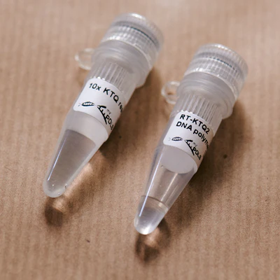
RNA into DNA and PCR in one step? Then, this enzyme will simplify PCR analysis from RNA templates reducing labor and time. RT-KTQ2 was evolved from the thermostable KlenTaq DNA polymerase with no significant reverse transcriptase activity. Four mutations ensure that the variant is reverse transcriptase active and even PCR active, while maintaining the thermostability. This allows to perform reactions at high temperatures minimizing problems encountered with strong secondary structures in RNA that melt at elevated temperatures. For further information refer to the original publication.
Detail
RNA into DNA and PCR in one step? Then, this enzyme will simplify PCR analysis from RNA templates reducing labor and time. RT-KTQ2 was evolved from the thermostable KlenTaq DNA polymerase with no significant reverse transcriptase activity. Four mutations ensure that the variant is reverse transcriptase active and even PCR active, while maintaining the thermostability. This allows to perform reactions at high temperatures minimizing problems encountered with strong secondary structures in RNA that melt at elevated temperatures. For further information refer to the original publication.
Available upon request and for R&D use only – Contact Us
RT-KTQ2 DNA polymerase is supplied as a 5 µM solution containing glycerol and is supplied together with 10x reaction buffer.
The enzyme can also be used for real-time cycling, when adding a suitable dye.
Other Products
HiDi® Taq DNA Polymerase
Product Info
Document
Product Info
HiDi® stands for High Discrimination of mismatches at the 3’-terminus of primers in PCR. This myPOLS Biotec enzyme family is optimized for this feature and is the first choice for applications that rely on this property such as allele-specific PCR (asPCR) or allele-specific amplification (ASA).Please note: This DNA polymerase is also available as a nuclease deficient variant, featuring higher robustness towards potential PCR inhibitors and compatibility with real-time dyes such as our GreenDye.Benchmarking with products of competitors conducted by us and others show that the HiDi® DNA polymerase family is the first choice for highly selective PCRs, such as genotyping by allele-specific PCR, HLA genotyping, analysis of single CpG methylation sites or the detection of mutations in a high background of wild-type sequences. By using HiDi® Taq DNA polymerase, less than 10 copies of a mutation can be detected in a background of >10.000 wild-type copies straight away without any other tedious assay optimization.HiDi® Taq DNA polymerase harbours a nuclease function and therefor is also suitable for use with hydrolysis probes (TaqMan® probes etc.). It has also been shown that HiDi® DNA polymerase family is highly suitable for quality control and mutation identification in CRISPR-Cas or TALEN-based applications.Several independently conducted studies show that HiDi® Taq DNA polymerase is ideally suited for use in asPCR in numerous research areas ranging from mutation detection to genome editing. (read more) For research use and further manufacturing.In case you are aiming to use our RUO products as components or for your development of e.g. an IVD medical device, please contact us.
Casestudies: HiDi® DNA Polymerase: Applications from mutation detection to genome editing (read more)
Example Primer Design
Matching vs. mismatching nucleotide is placed at the 3′-end of the primer for best discrimination results.
Example Results – There´s no accounting for taste
Cilantro: some people love it in their food, some hate it. Here we are detecting a genomic SNP (rs72921001) in HeLa genomic DNA. This SNP is reported to be close to a number of genes coding for olfactory receptors. (Reference: Eriksson N. et al. (2012), “A genetic variant near olfactory receptor genes influences cilantro preference.”)
Considering, that only the C-allele specific primer is extended and yielding in a specific amplicon, we can conclude a genetic predisposition in disliking cilantro, as this SNP is significantly associated with detecting a soapy taste to cilantro.
Allele-specific PCRs were performed from 1 ng/µl of HeLa gDNA in the presence of a realtime dye, indicating the amplification of the C-allele specific primer only. The A-allele specific primer is discriminated, thus not amplified up to 50 cycles.
PCR products were subsequently analysed on a 2.5% agarose gel. Specific product is visualized by ethidium bromide staining at the amplicon length of 109 bp.
Document
HiDi® stands for High Discrimination of mismatches at the 3’-terminus of primers in PCR. This myPOLS Biotec enzyme family is optimized for this feature and is the first choice for applications that rely on this property such as allele-specific PCR (asPCR) or allele-specific amplification (ASA).
Please note: This DNA polymerase is also available as a nuclease deficient variant, featuring higher robustness towards potential PCR inhibitors and compatibility with real-time dyes such as our GreenDye.
This product is suitable for rapid extraction of DNA from FFPE sample. This kit uses two combination methods. High-salt Bind is conducive to remove pigments or polysaccharides from complex FFPE samples, so as to improve the purity of nucleic acid and avoid blocking aligent 2100. Alcohol mediated adsorption is conducive to improve the nucleic acid yield of high-yield samples.
Details
Specifications
Features
Specifications
Main Functions
Isolation high pure total DNA from FFPE using high bind beads
Applications
PCR and viral DNA detection, etc.
Purification technology
Magnetic beads technology
Process method
Manual or automatic
Sample type
Paraffin embedded tissue samples
Sample amount
1-6 slices of 10-20μm
Elution volume
≥30μl
Time per run
≤60 minutes
Principle
This product is based on the purification method of high binding magnetic particles. The sample is lysed and digested under the action of lysate and Protease. DNA is released into the lysate. After adding magnetic particles and binding solution, DNA will be adsorbed on the surface of magnetic particles, and impurities such as proteins will be removed without adsorption. The adsorbed particles were washed with washing solution to remove proteins and impurities, washed with ethanol to remove salts, and finally DNA was eluted by Elution Buffer.
Advantages
High yield – most optimal process, recovery up to 90%
Economy – less than 50% of the price of Qiagen and other imported products
High purity – OD 260/280 : 1.7-1.9, OD 260/230 : 1.5-2.0
Kit Contents
Contents
D632301D
D632302D
Purification Times
48 Preps
96 Preps
MagPure Particles N
1.1 ml
2.5 ml
RNase A
10 mg
20 mg
Proteinase K
24 mg
48 mg
Protease Dissolve Buffer
3 ml
6 ml
Buffer DPS
60 ml
100 ml
Buffer ATL
15 ml
30 ml
Buffer BST1
30 ml
60 ml
Buffer BW1
13 ml
44 ml
Elution Buffer
15 ml
30 ml
Storage and Stability
Proteinase K, RNase A and MagPure Particles N should be stored at 2-8°C upon arrival. However, short-term storage (up to 12 weeks) at room temperature (15-25°C) does not affect their performance. The remaining kit components can be stored at room temperature (15-25°C) and are stable for at least 18 months under these conditions.
Document
This product is suitable for rapid extraction of DNA from FFPE sample. This kit uses two combination methods. High-salt Bind is conducive to remove pigments or polysaccharides from complex FFPE samples, so as to improve the purity of nucleic acid and avoid blocking aligent 2100. Alcohol mediated adsorption is conducive to improve the nucleic acid yield of high-yield samples.
Biotin-PEG3-alkyne is a biotin PEG reagent with an alkyne group that enables click reaction with azido molecules in the presence of Cu catalyst. The hydrophilic PEG spacer arm imparts water solubility that is transferred to the biotinylated molecule.
Document
Biotin-PEG3-alkyne is a biotin PEG reagent with an alkyne group that enables click reaction with azido molecules in the presence of Cu catalyst. The hydrophilic PEG spacer arm imparts water solubility that is transferred to the biotinylated molecule.