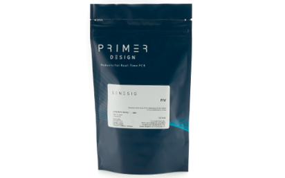
Exceptional value for money Rapid detection of all clinically relevant subtypes Positive copy number standard curve for quantification Highly specific detection profile High priming efficiency Broad dynamic detection range (>6 logs) Sensitive to < 100 copies of target
Accurate controls to confirm findings
Nucleic acid testing (NAT) is the method of choice for detection and quantification of a wide range of micro organisms. Primerdesign manufactures and supplies high quality quantitative real-time PCR kits for the detection and simultaneous quantification of numerous significant pathogens . A copy number standard curve is provided for quantification and an the internal extraction template (DNA or RNA), controls for the quality of the nucleic acid extraction and eliminates false negative results.
The kit is designed with the broadest possible detection profile to ensure that all clinically relevant strains and subtypes are detected. Target sequences are selected by working with data from key opinion leaders in the field. Multiple sequence alignments and unprecedented real-time PCR expertise in design and validation ensure the best possible kit.
Details of the target and priming specificity are included in the individual handbooks above.
Packaged, optimised and ready to use. Expect Better Data.
Q-PAGE™ Bis-Tris Precast Gel is a high-performance and easy to use precast polyacrylamide gel for electrophoresis in Bis-Tris buffer system (MOPS or MES). The optimized gel formula allows Q-PAGE™ Bis-Tris Precast Gel to show improved resolution, accurate results, and an extended shelf-life over conventional Laemmli Tris-HCl gels.
Q-PAGE™ Bis-Tris Precast Gels are available in gradient (4 to 12%) and fixed (8% and 12%) concentrations of polyacrylamide in 12-and 15-well formats. Two available cassette sizes, Mini (10 x 8.3 cm) and Midi (10 x 10 cm), are compatible with most popular protein electrophoresis systems. Q-PAGE™ Mini (QP2XXX) Gels are suitable for Bio-Rad® and other systems. Q-PAGE™ Midi (QP3XXX) Gels are suitable for Invitrogen® XCell SureLock® Mini-Cell, Invitrogen® Mini Gel Tank, Hoefer SE260, and other systems.
Key Features
User-friendly gel cassette:
Numbered and framed wells for sample loading
With cassette opener for easy use
Enhanced gel performance:
Enhanced band sharpness
Better resolution of small proteins
Stable for shipping at ambient temperature
Easy compatibility:
Available as homogeneous and adjusted gradient gels for a wide range of protein separation.
Compatible with most popular protein electrophoresis systems
Storage and stability
Store Q-PAGE™ Precast Gels at 4°C for periods up to 12 months.
Do not freeze Q-PAGE™ Precast Gels Remove tape and comb before electrophoresis.
Keep Q-PAGE™ Precast Gels flat during storage.
Document
Q-PAGE™ Bis-Tris Precast Gel is a high-performance and easy to use precast polyacrylamide gel for electrophoresis in Bis-Tris buffer system (MOPS or MES). The optimized gel formula allows Q-PAGE™ Bis-Tris Precast Gel to show improved resolution, accurate results, and an extended shelf-life over conventional Laemmli Tris-HCl gels.
Q-PAGE™ Bis-Tris Precast Gels are available in gradient (4 to 12%) and fixed (8% and 12%) concentrations of polyacrylamide in 12-and 15-well formats. Two available cassette sizes, Mini (10 x 8.3 cm) and Midi (10 x 10 cm), are compatible with most popular protein electrophoresis systems. Q-PAGE™ Mini (QP2XXX) Gels are suitable for Bio-Rad® and other systems. Q-PAGE™ Midi (QP3XXX) Gels are suitable for Invitrogen® XCell SureLock® Mini-Cell, Invitrogen® Mini Gel Tank, Hoefer SE260, and other systems.
Discover Huankai’s Modified MSRV Agar Base, designed for the selective detection and isolation of motile Salmonella species, ensuring reliable food and environmental testing.
Document
Discover Huankai’s Modified MSRV Agar Base, designed for the selective detection and isolation of motile Salmonella species, ensuring reliable food and environmental testing.
RAA uses a novel RNA substrate tagged with a fluorescent reporter molecule (fluor) on one end and a quencher on the other. In the absence of RNases, the physical proximity of the quencher dampens fluorescence from the fluor to extremely low levels. When RNases are present, however, the RNA substrate is cleaved, and the fluor and quencher are spatially separated in solution. This causes the fluor to emit a bright green signal when excited by light of the appropriate wavelength. Fluorescence can be readily detected with a fluorometer. Since the fluorescence of the RAA Substrate increases over time when RNase activity is present, results monitored with a fluorometer can be evaluated kinetically. The sequence of the RAA Substrate has been carefully optimized to detect several RNases, including RNase A, RNase T1, RNase I, micrococcal nuclease, S1 nuclease, mung bean nuclease, and Benzonase.
Document
RNase activity in a convenient and sensitive fluorimetric assay that delivers results in real time. Great for Quality Testing for RNase contamination of materials and supplies.