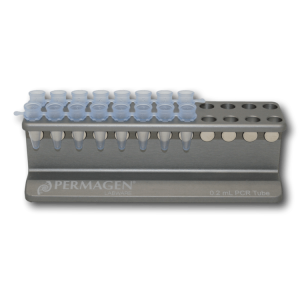
Permagen’s 0.2 mL PCR Strip Magnetic rack is designed for magnetic bead separations from Individual tubes, 8-strip, or 12-strip PCR tubes
Detail
Permagen’s 0.2 mL PCR Strip Magnetic rack is designed for magnetic bead separations from Individual tubes, 8-strip, or 12-strip PCR tubes
Accommodates any common PCR strips or individual PCR tubes
Tubes are angled and beads will be pulled to back wall allowing easy aspiration and tip tracking down the front wall of the tubes without disturbing bead pellet
Campylobacter jejuni is a curved, rod-shaped and microaerophillic gram negative bacterium. It is one of the most common causal agents of gastroenteritis with diarrhea as the main symptom. While infection of C. jejuni is seldom life-threatening, it is considered one of the most common food-borne bacteria with over 2 million people infected per year in US alone. Infection of C. jejuni usually results from consumption of poorly prepared food including undercooked meat (particularly poultry), untreated water or raw unpasteurized milk. Traditional identification of C. jejuni involves culturing, however the microaerophilic characteristic of this bacterium make the enrichment process laborious and costly.
Storage Conditions and Product Stability All kit components can be stored for 2 years after the date of production without showing any reduction in performance.
All kit components should be stored at -20°C upon arrival. Repeated thawing and freezing (> 2 x) of the Master Mix and Positive Control should be avoided, as this may affect the performance of the assay. If the reagents are to be used only intermittently, they should be frozen in aliquots.
This product is suitable for extracting total viral nucleic acid from cell-free/low-content cell biological samples such as body fluids, serums, plasma, soaking solutions, tissue homogenate supernatant, and culture supernatant. The Purified DNA/RNA is used for RT-PCR and PCR detection.
Details
Specifications
Features
Specifications
Main Functions
IVD5412 precast kit for MagMix 32, smart 32
Applications
RT-PCR,PCR,NGS
Products
Viral DNA / RNA, body cell DNA / RNA
Purification method
Polydisperse magnetic beads
Purification technology
Magnetic beads technolog
Process method
Manual or automatic
Sample type
Sample amount
200μl
Adaptive instrument
Nucleic acid extractor, pipetting workstation
Principle
This product is based on the purification method of high binding magnetic particles. The sample is lysed and digested under the action of lysate and Protease. DNA/RNA is released into the lysate. After adding magnetic particles and binding solution, DNA/RNA will be adsorbed on the surface of magnetic particles, and impurities such as proteins will be removed without adsorption. The adsorbed particles were washed with washing solution to remove proteins and impurities, washed with ethanol to remove salts, and finally DNA/RNA was eluted by Nuclease Free Water.
This kit is shipped and stored at room temperature and is valid for 12 months.
Document
This product is suitable for extracting total viral nucleic acid from cell-free/low-content cell biological samples such as body fluids, serums, plasma, soaking solutions, tissue homogenate supernatant, and culture supernatant. The Purified DNA/RNA is used for RT-PCR and PCR detection.
This ELISA kit is intended for the quantitative detection of IgG antibodies against Echinococcus multilocularis in human Serum.
This product is manufactured by Bordier Affinity Products in Switzerland and distributed in Germany exclusively by Milenia Biotec.
Method/Platform
ELISA in microplate format
Range/Assay Sensivity
pNPP, λ=405 nm
Test Principle
Specific antibodies in the sample bind to Echinococcus multilocularis Em2-Em18 antigens sensitized on microtiter plates. The presence of parasite specific antibodies is detected with a Protein A – alkaline phosphatase conjugate.