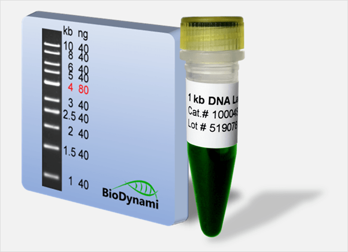
• For sizing and quantification of double strand DNA fragments. • Composed of ten bands as shown on right. • The 4 kb band with higher concentration is easily distinguishable from the others. • Premixed with 6X DNA loading buffer for direct gel loading.
Detail
1 kb DNA Ladder in 1% agarose gel
• For sizing and quantification of double strand DNA fragments. • Composed of ten bands as shown on right. • The 4 kb band with higher concentration is easily distinguishable from the others. • Premixed with 6X DNA loading buffer for direct gel loading.
CE-IVD marked version available for in vitro diagnostic use
Available in TaqMan format for analysis
Herpes Simplex Virus 2 (HSV-2) is a member of the herpes virus family, Herpesviridae. HSV-2 has a relatively large double-stranded DNA genome. HSVs are primarily transmitted by sexual intercourse, direct contact with lesions or perinatally. Most HSV positive cases are characterised by lesions on the skins and mucous membranes of the mouth and genitals. HSV infection can be either primary or a recurrence of a previous infection. More than 90% of the primary HSV infections are asymptomatic. Primary infection with HSV-1 can lead to gingivostomatitis, eczema herpeticum, keratoconjunctivitis and encephalitis. The primary symptoms of a secondary infection are skin lesions in the nose, mouth and genital regions. The infection is contagious, mainly during an epidemic.
HSV-2 TaqMan PCR Kit, 100 reactions
Ready to use format, including Master Mix for the target and PCR control to monitor for PCR inhibition and validate the quality
Specific Primer and Probe mix for the pathogen/virus/viroid of interest
Primer and Probe mix
Positive and negative control to confirm the integrity of the kit reagents
HSV-2 TaqMan PCR Probe/Primer Set and Controls, 100 reactions
Specific Primer/Probe mix and Positive Control for the pathogen/virus/viroid of interest
Nuclease-free water
Can be used together with Norgen’s PCR Master Mix (#28007) or customer supplied master mix
Storage Conditions and Product Stability All kit components can be stored for 2 years after the date of production without showing any reduction in performance.
All kit components should be stored at -20°C upon arrival. Repeated thawing and freezing (> 2 x) of the Master Mix and Positive Control should be avoided, as this may affect the performance of the assay. If the reagents are to be used only intermittently, they should be frozen in aliquots.
An enhanced PCR master mix for allele-specific assays. Improved signal to noise ratio and tight clustering. Developed specifically for genotyping direct from crude DNA samples.
PACE 2.0 Genotyping Master Mix ensures an unrivalled signal-to-noise ratio and produces tight data clusters, even when working with high-throughput, crude DNA preps, resulting in consistently exceptional performance. Efficiently streamline your workflow and reduce costs without compromising the quality of your results.
PACE 2.0 Genotyping Master Mix is an ideal solution for challenging starting material. PACE 2.0 has been specially formulated to overcome the obstacles presented by common PCR inhibitor compounds, such as phenols and tannins. Even notoriously tricky samples like oil palm and conifers can still be assayed using hot shot or other crude DNA prep methods and deliver reliable and accurate data.
PACE 2.0 Genotyping Master Mix uses a novel, universal, fluorescent reporting cassette to produce machine-readable fluorescent signals corresponding to genotypes. PACE 2.0 compatible genotyping assays are comprised of two competitive allele-specific forward primers (which differ in their terminal 3’ bases and unique 5’ tail sequences) and a common, reverse primer. PACE 2.0 Genotyping Master Mix is supplied with ROX normalising dye at a range of levels to ensure compatibility with your qPCR instrument or reader.
Genotyping assay designs are available from 3CR Bioscience through our free assay-design service; once designed, users can purchase assay primers independently or through 3CR Bioscience using our partial or full-assay validation service. PACE 2.0 Genotyping Master Mix is also compatible with KASP™ and Amplifluor® marker assays.
REQUIRED COMPONENTS
qPCR machine or Thermocycler + Fluorescent plate reader
PCR plate or equivalent and appropriate optically clear seal
1 Layer Cell Factory One Wide Mouth And One Narrow Mouth
Product Info
Document
Product Info
1 Layer Cell Factory One Wide Mouth And One Narrow Mouth
Huayi Cell Factory is a robust and easy-to-use cell culture platform for applications in the production of human and animal vaccines, therapeutic proteins, cell therapy, and gene therapy.
To address the needs of your workflow, We have three kinds of mouth, which is available on most cell culture products to ensure consistent performance from lot to lot and from format to format.
The product assembled with ultrasonic welded technology
Versatile port design facilitates both pouring and aseptic filling techniques
Gamma radiation sterilization
the cell culture surface area
of one 10-layer Cell Factory unit is equivalent to the area
of 36 T-175 flasks
Document
Huayi Cell Factory is a robust and easy-to-use cell culture platform for applications in the production of human and animal vaccines, therapeutic proteins, cell therapy, and gene therapy.
To address the needs of your workflow, We have three kinds of mouth, which is available on most cell culture products to ensure consistent performance from lot to lot and from format to format.