Fluorescent Universal Human IgG/IgM Lateral Flow Serology Kit (Quantum Dots)
Facebook
X
Pinterest
Email
Antibody tests are a method of choice to determine if a person has been exposed to a pathogen or not. They are also incredibly valuable in the detection of autoantibodies that can be found in human autoimmune disorders. In this test, a biotinylated antigen (User supplied) is mixed with a biotinylated rabbit IgG (bind to goat anti rabbit control line) and sample (human sera or plasma) is simply mixed into with the specially designed assay running buffer in a well of the supplied 96-well plate, mixed and is then added to the sample port of the cassette. Generally, the reaction is complete in 10-15 minutes. It is very important to note that the relative stoichiometry between the biotinylated antigen, biotinylated rabbit IgG added, and the streptavidin gold is critical for assay optimization. The appropriate concentration of biotinylated antigen to use with strips is dependent upon the purity and sequence and a standard curve can be used to determine the relative ratio (generally between 1ng-100ng per test). A positive control line (biotin-rabbit IgG) antibody will bind to the goat anti rabbit (GAR) line on the test to ensure the assay is running appropriately.
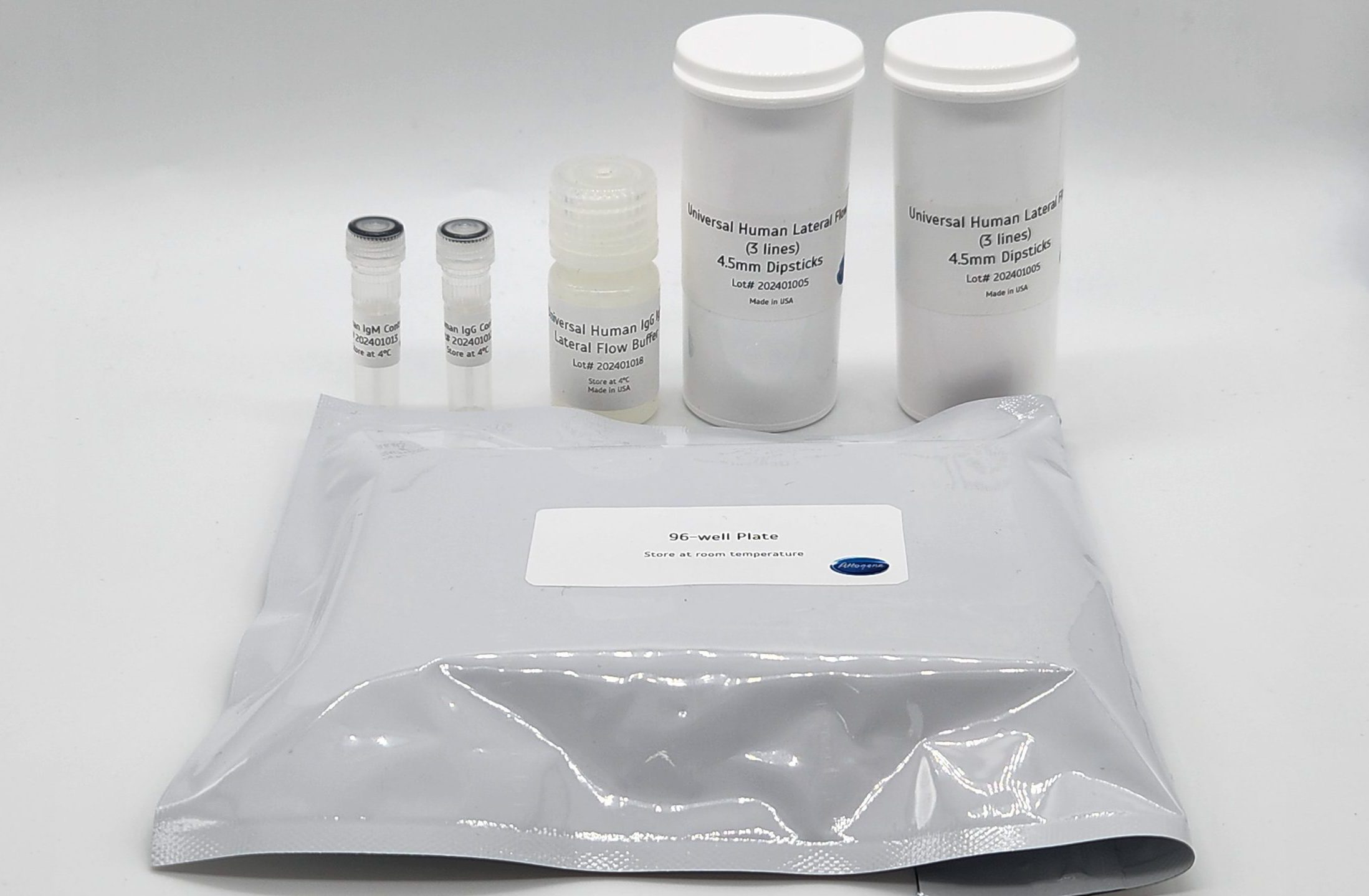
Attogene’s Human IgG/IgM universal fluorescent lateral flow assay kit is a ready-to-use, universal test strip, which is based on the lateral flow technology that uses 655nm Emission Quantum Dot particles containing streptavidin to conveniently capture biotinylated antigens. The device is designed to easily develop qualitative or quantitative rapid test systems for detection of anti-human IgG and IgM antibody that react to the any antigen that can be biotinylated (i.e. viral antigen, autoimmune antigen) and is easily customizable providing every laboratory with the possibility to perform assay feasibility.
Formats (fluorescent broad range UV light excitation range of 300nm to 400nm, 610nm emission) Streptavidin conjugate pad):
Other Products
IST-104 PeelASeal FoilTM Heat Sealing Film
Product Info
Document
Product Info
Overview
Peelable heat sealing foil which is suitable for low temperature storage, high temperature uses and PCR.
Heat sealing offers a 100% effective method of plate sealing, for complete seal integrity, as well as being quick and cost effective
Our PeelASeal Foil is a laminate seal compatible with polypropylene plates
It can be removed from polypropylene plates by peeling, even with a plate which has been removed directly from -80 °C storage
PeelASeal Foil forms a complete seal to a plate enabling very low temperature uses, including very low temperature storage, and high temperature uses, such as PCR (when used with a pressurized heated lid)
The seal demonstrates moderate solvent resistance and can be utilized for short term compound storage at room temperature
The seal is available as sheets, for use with manual and semi-automated sealers, such as our HeatASeal 500 Sealing Machine
Also available in multiple roll formats compatible with specified automated heat sealers, such as our Wasp or Chameleon XT
Document
Peelable heat sealing foil which is suitable for low temperature storage, high temperature uses and PCR.
TCO-PEG12-DBCO is a long chain PEGreagent containing a TCO and a DBCO moiety. TCO group specifically and efficiently reacts with terrahydrazine under mild condition. DBCO is very reactive toward Azide through click chemistry, the PEG spacer increases the aqueous solubility of the reagent. Reagent grade, for research purpose. Please contact us for GMP-grade inquiries.
Document
TCO-PEG12-DBCO is a long chain PEGreagent containing a TCO and a DBCO moiety. TCO group specifically and efficiently reacts with terrahydrazine under mild condition. DBCO is very reactive toward Azide through click chemistry, the PEG spacer increases the aqueous solubility of the reagent. Reagent grade, for research purpose. Please contact us for GMP-grade inquiries.
RNA/microRNA/DNA/Proteins are preserved for more than 2 years at room temperature in Norgen’s Urine Preservative
Compatible with most DNA, Total RNA, microRNA and protein isolation methods
Preservative is available in a single dose liquid format (ampule)
Preservative is also available in a dried format in tubes – Urine Collection and Preservation Tubes
Urine Collection and Preservation Devices are available in 4 convenient sizes: 5 cc tubes, 15 cc tubes, 50 cc tubes, and 120 cc cups
Norgen’s Urine Preservative is designed for the rapid preservation of DNA, RNA, microRNA and proteins from fresh urine samples. In addition, the Urine Preservative eliminates the need to immediately process or freeze samples and allows the samples to be shipped to centralized testing facilities at ambient temperatures. The components of the Urine Preservative allow samples to be stored for over 2 years at room temperature with no detected degradation of urine DNA, RNA or proteins.
Norgen’s Urine Preservative is available in 2 convenient formats:
1. Urine Preservative Single Dose Ampules With this product Norgen’s Urine Preservative is supplied in a liquid format in single dose ampules. The user simply collects 5 – 50 mL of urine into a urine collection container and adds the contents of the Urine Preservative Single Dose (Cat# 18126). The urine and preservative are then mixed, and the urine nucleic acids and proteins are preserved at room temperature.
2. Urine Collection and Preservation Tubes Norgen’s Urine Preservative is also available in a dried format in Norgen’s Urine Collection and Preservation Tubes. The user simply collects urine into the tubes and mixes gently until the orange preservative pellet in the tube has dissolved. Norgen’s Urine Collection and Preservation Tubes are available in 3 convenient sizes:
Select Urine Tested with the Norgen Urine Collection and Preservation Tubes
Human Mouse Lynx Wolf Urine collected from snow
Available Sizes and Formats
Urine Collection and Preservation Tubes (50 cc) – Urine inputs from 5 – 45 mL
Storage Conditions and Product Stability All tubes should be kept tightly sealed and stored at room temperature (15 – 25°C) for up to 2 years without any reduction in performance.