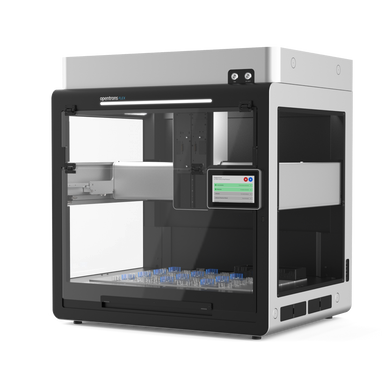
The Opentrons Flex HEPA/UV Module enables you to run sensitive contamination-prone applications. It removes 99.99% of 0.3 μm DNA-containing particulates and biological contaminants like bacteria, fungi, and other microorganisms from the air, creating a clean work environment inside the Opentrons Flex.
Detail
The Opentrons Flex HEPA/UV Module enables you to run sensitive contamination-prone applications. It removes 99.99% of 0.3 μm DNA-containing particulates and biological contaminants like bacteria, fungi, and other microorganisms from the air, creating a clean work environment inside the Opentrons Flex.
Other Products
D3187 HiPure HP Plant DNA Kit
Product Info
Document
Product Info
Introduction
HiPure Plant DNA Mini Kit supplies a simple and rapid extraction of genomic DNA from different plant samples. The kit is based on silica gel column and CTAB lysis purification technology. The whole extraction process is only 30-50 minutes. Purified DNA can be used directly for PCR, SSR, AFLP, RAPD and Southern Blot, etc.
Details
Specifications
Features
Specifications
Main Functions
Isolation total DNA from 150 mg plant and fungal tissue
This product is based on silica column purification. The sample is lysed with CATB Buffer. DNA is released into the lysate. Cell debris, precipitated proteins and polysaccharides are removed by chloroform extraction. After adjust the binding condition, transfer to an adsorption column. DNA is adsorbed on the membrane, while protein is not adsorbed and is removed with filtration. After washing proteins and other impurities, nucleic acid was finally eluted with low-salt buffer (10mm Tris, pH9.0, 0.5mm EDTA).
Advantages
Broad spectrum – suitable for extracting DNA from various plant samples
Fast – several samples can be extracted in 30 minutes by silica technology
Good repeatability – silica gel column purification technology can obtain ideal results every time
Kit Contents
Contents
D318702
D318703
Purification Times
50 Preps
250 Preps
Buffer PAL
60 ml
200 ml
Buffer GWP
60 ml
200 ml
Buffer DW1
30 ml
150 ml
Buffer GW2*
20 ml
50 ml
Buffer AE
20 ml
60 ml
HiPure DNA Mini Columns II
50
250
2 ml Collection Tubes
50
250
Storage and Stability
This product can be stored dry at room temperature (15-25°C) and are stable for at least 18 months under these conditions.
HiPure Plant DNA Mini Kit supplies a simple and rapid extraction of genomic DNA from different plant samples. The kit is based on silica gel column and CTAB lysis purification technology. The whole extraction process is only 30-50 minutes. Purified DNA can be used directly for PCR, SSR, AFLP, RAPD and Southern Blot, etc.
TCO-PEG6-DBCO is a PEG6 linker featuring a trans-cyclooctene and a DBCO group. DBCO is a click chemistry handle which easily reacts with azides, while the TCO function readily reacts with tetrazine groups in target compounds.
Document
TCO-PEG6-DBCO is a PEG6 linker featuring a trans-cyclooctene and a DBCO group. DBCO is a click chemistry handle which easily reacts with azides, while the TCO function readily reacts with tetrazine groups in target compounds.
[DL4000] ExcelDye™ 6X DNA Loading Dye, Tri-color, 5 ml x 2
Product Info
Document
Product Info
Description
The ExcelDye™ 6× DNA Loading Dye (Tri-Color) is pre-mixed buffer for tracking the DNA sample during the electrophoresis on agarose or polyacrylamide gels. It contains three dyes (Xylene cyanol FF, Bromophenol blue, Orange G) for tracking the DNA migration. The Xylene cyanol FF, Bromophenol blue and Orange G migrate at approximately 800 bp, 150 bp and 30 bp on a standard 2% TAE agarose gel respectively (4,000 bp, 500 bp and 50 bp on 1% TAE agarose gel respectively). The included glycerol keeps the DNA at the bottom of the well and the presence of EDTA chelates divalent metal ions to prevent the process of metal-dependent nuclease.
Composition
0.03% Xylene cyanol FF
0.03% Bromophenol blue
0.15% Orange G
10 mM Tris-HCl (pH 8.0)
60% glycerol
60 mM EDTA
Storage
4°C for 12 months -20°C for 36 months
Document
The ExcelDye™ 6× DNA Loading Dye (Tri-Color) is pre-mixed buffer for tracking the DNA sample during the electrophoresis on agarose or polyacrylamide gels. It contains three dyes (Xylene cyanol FF, Bromophenol blue, Orange G) for tracking the DNA migration. The Xylene cyanol FF, Bromophenol blue and Orange G migrate at approximately 800 bp, 150 bp and 30 bp on a standard 2% TAE agarose gel respectively (4,000 bp, 500 bp and 50 bp on 1% TAE agarose gel respectively). The included glycerol keeps the DNA at the bottom of the well and the presence of EDTA chelates divalent metal ions to prevent the process of metal-dependent nuclease.