An enhanced PCR master mix for allele-specific assays. Improved signal to noise ratio and tight clustering. Developed specifically for genotyping direct from crude DNA samples.
PACE 2.0 Genotyping Master Mix ensures an unrivalled signal-to-noise ratio and produces tight data clusters, even when working with high-throughput, crude DNA preps, resulting in consistently exceptional performance. Efficiently streamline your workflow and reduce costs without compromising the quality of your results.
PACE 2.0 Genotyping Master Mix is an ideal solution for challenging starting material. PACE 2.0 has been specially formulated to overcome the obstacles presented by common PCR inhibitor compounds, such as phenols and tannins. Even notoriously tricky samples like oil palm and conifers can still be assayed using hot shot or other crude DNA prep methods and deliver reliable and accurate data.
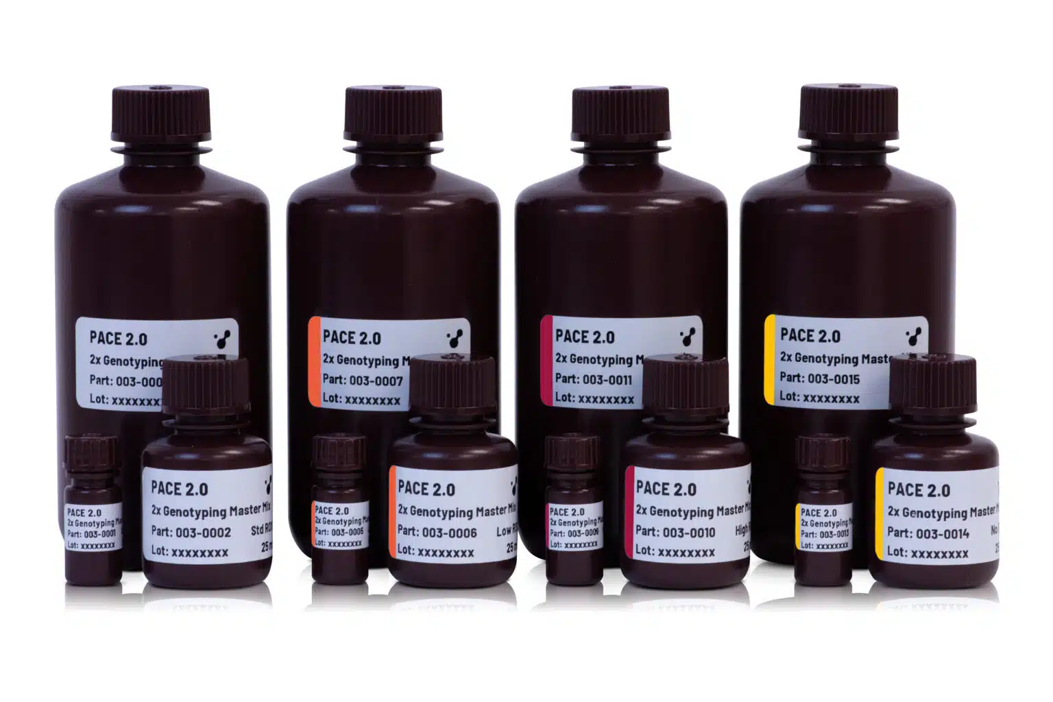
PACE 2.0 Genotyping Master Mix uses a novel, universal, fluorescent reporting cassette to produce machine-readable fluorescent signals corresponding to genotypes. PACE 2.0 compatible genotyping assays are comprised of two competitive allele-specific forward primers (which differ in their terminal 3’ bases and unique 5’ tail sequences) and a common, reverse primer. PACE 2.0 Genotyping Master Mix is supplied with ROX normalising dye at a range of levels to ensure compatibility with your qPCR instrument or reader.
Genotyping assay designs are available from 3CR Bioscience through our free assay-design service; once designed, users can purchase assay primers independently or through 3CR Bioscience using our partial or full-assay validation service. PACE 2.0 Genotyping Master Mix is also compatible with KASP™ and Amplifluor® marker assays.
REQUIRED COMPONENTS
qPCR machine or Thermocycler + Fluorescent plate reader
PCR plate or equivalent and appropriate optically clear seal
Template DNA
PCR-grade water
Genotyping assays
Other Products
Cryptococcus neoformans TaqMan PCR Detection Kits
Product Info
Document
Product Info
Overview
Detection kits for Cryptococcus neoformans
Available in TaqMan format for analysis
Cryptococcus neoformans is an encapsulated yeast. Infection with C. neoformans is known as cryptococcosis and is the cause of the most common life-threatening meningitis in patients with weakened immune systems, particularly in advanced HIV/AIDS. C. neoformans is commonly found in soil throughout the world. Human infection of C. neoformans occurs via inhalation of aerosolized spores. From the lungs, C. neoformans is spread hematogenously to the Central Nervous System (CNS), resulting in meningoencephalitis. Although the availability of antiretroviral therapy in the developed world has reduced the incidents of cryptococcosis, it is still a major problem in developing countries and is one of the leading causes of death in patients with HIV/AIDS. In fact, one of the major challenges in treating cryptococcosis is that many patients with cryptococcal CNS disease are asymptomatic in terms of cryptococcal pneumonia, making it difficult for early detection.
Storage Conditions and Product Stability All kit components can be stored for 2 years after the date of production without showing any reduction in performance.
All kit components should be stored at -20°C upon arrival. Repeated thawing and freezing (> 2 x) of the Master Mix and Positive Control should be avoided, as this may affect the performance of the assay. If the reagents are to be used only intermittently, they should be frozen in aliquots.
Our SNPsig® kits use our own proprietary genotyping method to enable the identification of SARS-CoV-2 variants of concern. These products can be used on any real-time PCR machine using familiar protocols, whilst resulting in exceptional genotyping data.
Positive control templates for wild-type and variants are supplied in every kit to make data interpretation simple.
SNPsig® EscapePLEX™ SARS-CoV-2 also incorporates the two gene (ORF1ab and M Gene) assay to provide a confirmatory detection of SARS-CoV-2.
DETECTION PROFILE: The E484K, K417N, K417T and P681R mutations which are found in Beta, Gamma, Delta and Delta Plus Variants of Concern
For general laboratory and research use only.
Document
Rapid detection of specific detection profiles High priming efficiency Sensitive to < 100 copies of target
Positive copy number standard curve for quantification
Accurate controls to confirm findings
96 reactions
Our Blyscan™ Glycosaminoglycan Kit has been a ‘go-to’ Solution for reliable sGAG and Proteoglycan Analysis for many years! Blyscan utilises a dye-binding approach to quantitatively measure sulfated glycosaminoglycans (sGAG) and proteoglycans in cells, tissues and fluids from a wide range of in-vivo and in-vitro sources.
Colorimetric Detection (656nm) (Endpoint)
Understanding Glycosaminoglycans (GAGs) and Proteoglycans
Glycosaminoglycans (GAGs) are a type of negatively charged polysaccharide that play crucial roles in various biological processes. They are composed of repeated disaccharide units, typically of N-acetylated or N-sulfated hexosamine paired with a uronic acid (GlcA or IdoA) or galactose. Sulfate groups can also be added to give sulfated GAGs an overall negative charge that influences cell interactions and also enable binding by our Blyscan dye reagent.
Common examples of GAGs include Chondroitin Sulfate, Dermatan Sulfate, Heparin, Heparan Sulfate, and Keratan Sulfate. Note that Hyaluronic Acid is a non-sulfated GAG and cannot be detected by the Blyscan assay. If you need to measure hyaluronic acid instead, we recommend using our Purple-Jelley kit!
The Role of Glycosaminoglycans in Tissues
GAGs and proteoglycans have essential functions in tissues and organisms, providing biophysical support through scaffolding and maintaining cartilage hydration. They also play a vital role in biochemical processes such as cell adhesion and signalling.
What is the origin of the Blyscan assay name?
Blyscan is an Old English word meaning ‘to shine’ and from which the word ‘blush’, (blushing), may have been derived. This was an appropriate choice as the Blyscan Assay contains a blue dye which ‘blushes’ bright pink when it binds to sulphated glycosaminoglycans!
How does the Blyscan assay work?
Step 1. Blyscan dye reagent contains DMMB dye in an optimised buffer. Addition of Dye reagent to samples containing sGAG results in the formation of a dye/sGAG complex due to a charge interaction between dye and GAG sulfate groups.
Step 2. Over a 30 minute incubation Dye-labelled sGAGs precipitate out of solution and are collected by centrifugation. Following removal of unbound dye, the remaining bound dye is released from the complex by addition of dye dissociation reagent. Released dye is quantified spectrophotometrically.
Step 3. The sGAG content of unknown samples may be quantified by comparison against a calibration curve prepared using a standard of purified Chondroitin-4-sulfate supplied with the kit.
A list of suggested sample types can be found under the ‘Assay Specification‘ tab.
The Blyscan Dye reagent is formulated to miminise binding to other charged sample components such as nucleic acids, a problem with some older dye-based sGAG assays.
Assay range
2.5 – 50µg/ml
Limit of Detection
2.5µg/ml
Detection Method
Colorimetric Detection (656nm) (Endpoint)
Measurements per kit
110 in total (allows a maximum of 48 samples to be run in duplicate alongside a standard curve).
In-vivo: Liquid samples, including fluids such as urine, amniotic or synovial fluid.
In-vitro: Solid samples, such as deposited ECM on 2D/3D culture surfaces.by enzymatic treatment
In-vivo: Liquid samples, Culture media during 2D/3D cell culture.
The assay requires that sulfated polysaccahrides or sGAGs are in a soluble form. A preliminary enzymatic extraction step is required for solid samples (enzyme not supplied with kit).
The assay is not suitable for use with samples containing alginates or that comprise degraded sulfated disaccharide fragments.
Precautions
This kit is designed for research use only. Not for use in diagnostic procedures. Kit requires access to a centrifuge, as well as a spectrophotometer/colorimeter capable of absorbance detection at 656nm. Specific sample preparation protocols may require customer to provide further reagents, consult assay manual for further information.
Blyscan sGAG kit contents:
1. Blyscan Dye Reagent (1x110ml)
2.sGAG Reference Standard (1x5ml, 100µg/ml Bovine tracheal chondroitin 4-sulfate)
3. Dissociation Reagent (1x110ml)
4. Sodium Nitrite (1x15ml)
5. Acetic Acid (1x15ml)
6. Ammonium Sulfamate (1x15ml)
7. 1.5ml micro-centrifuge tubes for dye-labelling reaction.
8. Assay kit manual
NB: Additional reagents may be required for sample preparation prior to assay. Consult manual or contact us for further details.
Document
Our Blyscan™ Glycosaminoglycan Kit has been a ‘go-to’ Solution for reliable sGAG and Proteoglycan Analysis for many years! Blyscan utilises a dye-binding approach to quantitatively measure sulfated glycosaminoglycans (sGAG) and proteoglycans in cells, tissues and fluids from a wide range of in-vivo and in-vitro sources. Colorimetric Detection (656nm) (Endpoin