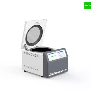
Key features: 1. High-definition LCD touch screen, simultaneously displaying operational parameters: speed, temperature, vacuum, and running time. 2. Delayed start: after reaching a fixed speed, Start the vacuum pump to effectively prevent sample mixing. 3. Fully automatic vacuum control system, vacuum setting range0.1~1000mbar, Control accuracy0.1mbar. 4. 1.5/2ml Rotor stacking for simultaneous use; rotor replacement without tools. 5. Programmable with temperature and pressure settings; capable of storing 36 programs 6. Compatible with various rotor heads; transparent glass window for easy observation. 7. Magnetic drive: maintenance-free, non-contact drive, fully sealed. 8. Safety in use: inductive electric self-suction door lock ensures the centrifugal concentrator can only start when the door cover is fully closed and locked. 9. Can be used in conjunction with freeze-drying systems Model: SD-25/SD-25C
Brand: Yingtai
Detail
SD-25/SD-25C Vacuum Centrifugal Concentrator
Ambient temperature type/Refrigerated type
Technical Parameter
Temperature range
Room Temperature~+80℃
-10℃~+80℃
Temperature Accuracy
士1℃
Max.Speed
2500rpm
Max.Capacity
6x100ml
Timer
0~99h59min
Vacuum control range
0.1~1000mbar
Main unit withstands vacuum levels
0.01mbar
Noise
≤55dBA
≤65dBA
Voltage
0.7kw
1.3kw
Power supply
220v,50Hz
Dimension(L×W×Hmm)
360*448*304mm
440*653*420mm
Adapter Rotor
Rotor Type
Rotor Volume
Rotor Type
Rotor Volume
Angle Rotor
42×1.5/2ml
Angle Rotor
8×50ml
Angle Rotor
108×1.5/2ml
Angle Rotor
6×100ml
Angle Rotor
48×5ml
Angle Rotor
16*20ml Sample vial
Angle Rotor
30×10ml
Angle Rotor
16*5ml+8*4ml+8*3ml
Angle Rotor
24×15ml
Microplate Rotor
2×96well
Other Products
Fecal DNA Collection & Preservation Mini Tubes
Product Info
Document
Product Info
Overview
No need to immediately process samples
Includes pre-loaded beads for effective sample homogenization by bead beating (tubes are fully compatible with commercially available bead beating systems)
DNA preservation at room temperature over 2 years
Ship fecal samples at room temperature safely
Compatible with most DNA isolation methods
Norgen’s Fecal DNA Collection & Preservation Mini Tubes are designed for the rapid preservation of DNA from fresh fecal specimens. These tubes contain Norgen’s Fecal DNA Preservative in a liquid format. Moreover, each collection tube is preloaded with beads for effective sample homogenization in any commercially available bead beater equipment (e.g. MP Biomedicals’ FastPrep®-24). The user simply collects fecal samples into the tubes and mixes by shaking or vortexing until the feces is mixed well with preservative. The preservative prevents the growth of Gram-negative and Gram-positive bacteria and fungi, and also inactivates viruses allowing the resulting non-infectious samples to be handled and shipped safely. In addition, the preservative eliminates the need to immediately process or freeze samples and allows the samples to be shipped to centralized testing facilities at ambient temperatures. The components of the preservative allow the DNA in the samples to be preserved at room temperature for over 2 years.
This kit is highly suited to collection and preservation of fecal material from mouse, hamster, chinchilla, rabbit and more for microbiome studies and other applications.
DNA Isolation from Preservative
Fecal samples collected and preserved in Norgen’s Fecal DNA Collection and Preservation Mini Tubes are compatible with most fecal DNA isolation methods. Samples stored in the tubes have been used successfully with all of Norgen Biotek’s stool-based DNA isolation kits (please refer to Related Products table) and reagents.
Free Download
Extracting Biological Insights from Stool
Tips and tricks for isolating high yield and quality DNA, RNA, miRNA and EV’s from fecal samplesDownload for Free
Storage Conditions and Product Stability All tubes should be kept tightly sealed and stored at room temperature (15 – 25°C) for up to 2 years without any reduction in performance.
Kit Components
Cat. 27650
Fecal DNA Collection & Preservation Mini Tubes (containing liquid preservative and beads)
Short term stability: 2-8oC, Long term stability: See individual component labels
Stability:
> 2 years under recommended storage conditions
Analyte:
Glucose Oxidase
Assay Format:
Spectrophotometer, Microplate, Auto-analyser
Detection Method:
Absorbance
Wavelength (nm):
510
Signal Response:
Increase
Linear Range:
0.01 to 0.08 U/mL of glucose oxidase per assay
Limit of Detection:
10 U/L
Reaction Time (min):
~ 20 min
Application examples:
Enzyme preparations, and other materials (e.g. biological cultures, samples, etc.).
Method recognition:
Novel method
The Glucose Oxidase assay kit is a simple procedure for the rapid and reliable measurement and analysis of glucose oxidase activity in industrial enzyme preparations and bread improver mixtures.
All reagents stable for > 12 months after preparation
Simple format
Mega-Calc™ software tool is available from our website for hassle-free raw data processing
Standard included
Suitable for manual, microplate and auto-analyser forma
Document
The Glucose Oxidase assay kit is a simple procedure for the rapid and reliable measurement and analysis of glucose oxidase activity in industrial enzyme preparations and bread improver mixtures.
Aspergillus niger Nucleic acid testing (NAT) is the method of choice for detection and quantification of a wide range of micro organisms. Primerdesign manufactures and supplies high quality quantitative real-time PCR kits for the detection and simultaneous quantification of numerous significant pathogens . A copy number standard curve is provided for quantification and an the internal extraction template (DNA or RNA), controls for the quality of the nucleic acid extraction and eliminates false negative results.
The kit is designed with the broadest possible detection profile to ensure that all clinically relevant strains and subtypes are detected. Target sequences are selected by working with data from key opinion leaders in the field. Multiple sequence alignments and unprecedented real-time PCR expertise in design and validation ensure the best possible kit.
Details of the target and priming specificity are included in the individual handbooks above.
Packaged, optimised and ready to use. Expect Better Data.
Document
Exceptional value for money Rapid detection of all clinically relevant subtypes Positive copy number standard curve for quantification Highly specific detection profile High priming efficiency Broad dynamic detection range (>6 logs) Sensitive to < 100 copies of target
Accurate controls to confirm findings