Streptavidin Europium Chelate Microspheres For Lateral Flow
Facebook
X
Pinterest
Email
Europium Chelate Microspheres Nanoparticle Streptavidin Conjugate for Lateral Flow
Our Streptavidin Europium Chelate Microspheres conjugate is manufactured using special conjugation technology and functonally tested by lateral flow. We have coated our high quality nanoparticles with a proprietary surface coat that covalently binds the biotin forming ultra stable conjugates. The resulting Streptavidin Europium Chelate Microspheres can irreversibly bind biotin.
Detail
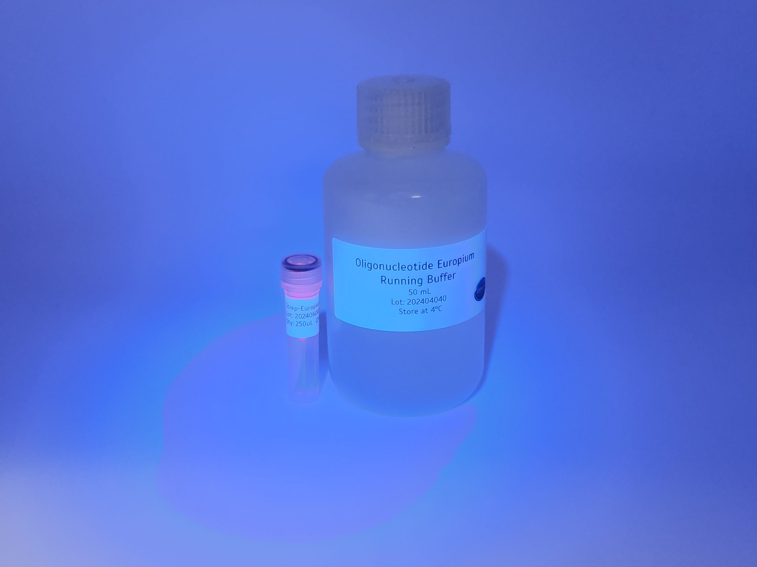
Ready to Use Streptavidin Europium Chelate Microspheres
Europium Chelate Microspheres Nanoparticle Streptavidin Conjugate for Lateral Flow
Our Streptavidin Europium Chelate Microspheresconjugate is manufactured using special conjugation technology and functonally tested by lateral flow. We have coated our high quality nanoparticles with a proprietary surface coat that covalently binds the biotin forming ultra stable conjugates. The resulting Streptavidin Europium Chelate Microspheres can irreversibly bind biotin.
Europium dyed polystyrene beads conjugated to streptavidin
250ul of a 1mg/ml stock ready to use for Lateral Flow
50mL Lateral FLow Running Buffer designed for Streptavidin Europium Chelate Microspheres
N-t-Boc-Aminooxy-PEG4-N-(PEG2-Propargyl) is a click chemistry crosslinker. The propargyl group is reactive with azide-containing compounds or biomolecules through copper catalyzed Click Chemistry to yield a stable triazole linkage. t-Boc-aminooxy can be deprotected under mild acidic conditions. The hydrophilic PEG linker improves solubility in aqueous media.
Document
N-t-Boc-Aminooxy-PEG4-N-(PEG2-Propargyl) is a click chemistry crosslinker. The propargyl group is reactive with azide-containing compounds or biomolecules through copper catalyzed Click Chemistry to yield a stable triazole linkage. t-Boc-aminooxy can be deprotected under mild acidic conditions. The hydrophilic PEG linker improves solubility in aqueous media.
Short term stability: 2-8oC, Long term stability: See individual component labels
Stability:
> 2 years under recommended storage conditions
Analyte:
L-Fucose
Assay Format:
Spectrophotometer, Microplate, Auto-analyser
Detection Method:
Absorbance
Wavelength (nm):
340
Signal Response:
Increase
Linear Range:
0.5 to 100 µg of L-fucose per assay
Limit of Detection:
0.68 mg/L
Reaction Time (min):
~ 10 min
Application examples:
L-Fucose is present as the main component in fucoidan (a marine polysaccharide), foods, pharmaceuticals and other materials (e.g. biological samples, etc.).
Method recognition:
Novel method
The L-Fucose test kit is a simple, rapid and reliable method, for the measurement and analysis of L-Fucose in plant extracts, biological samples and other materials. This kit can be used in the measurement of α-fucosidases that do not act on chromogenic substrates.
Note for Content: The number of manual tests per kit can be doubled if all volumes are halved. This can be readily accommodated using the MegaQuantTM Wave Spectrophotometer (D-MQWAVE).
All reagents stable for > 2 years after preparation
Only enzymatic kit available
Simple format
Rapid reaction time (~ 10 min)
Mega-Calc™ software tool is available from our website for hassle-free raw data processing
Standard included
Suitable for manual, microplate and auto-analyser formats
Document
The L-Fucose test kit is a simple, rapid and reliable method, for the measurement and analysis of L-Fucose in plant extracts, biological samples and other materials. This kit can be used in the measurement of α-fucosidases that do not act on chromogenic substrates.
DBCO-PEG4-triethoxysilane is a PEG linker containing a triethoxysilane moiety and a DBCO group. Triethoxysilane is commonly used for surface modifications. DBCO group can react with azide-bearing compounds or biomolecules to form a stable triazole linkage without copper catalyst. The hydrophilic PEG chain increasse the water solubility of a compound in aqueous media. Reagent grade, for research purpose. Please contact us for GMP-grade inquiries.
Document
DBCO-PEG4-triethoxysilane is a PEG linker containing a triethoxysilane moiety and a DBCO group. Triethoxysilane is commonly used for surface modifications. DBCO group can react with azide-bearing compounds or biomolecules to form a stable triazole linkage without copper catalyst. The hydrophilic PEG chain increasse the water solubility of a compound in aqueous media. Reagent grade, for research purpose. Please contact us for GMP-grade inquiries.