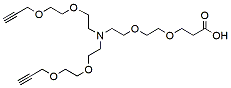
N-(Acid-PEG2)-N-bis(PEG2-propargyl) is reactive with azide-bearing compounds or biomolecules via copper catalyzed azide-alkyne Click Chemistry to yield a stable triazole linkage. The terminal carboxylic acid can react with primary amino groups in the presence of activators (e.g. EDC, or HATU) to form a stable amide bond. Reagent grade, for research purpose. Please contact us for GMP-grade inquiries.
Detail
N-(Acid-PEG2)-N-bis(PEG2-propargyl) is reactive with azide-bearing compounds or biomolecules via copper catalyzed azide-alkyne Click Chemistry to yield a stable triazole linkage. The terminal carboxylic acid can react with primary amino groups in the presence of activators (e.g. EDC, or HATU) to form a stable amide bond. Reagent grade, for research purpose. Please contact us for GMP-grade inquiries.
2-(Propargyl-PEG4-amido)-1,3bis(PEG1-methyl ester) is a crosslinker that can react with azide compounds or biomolecules via copper catalyzed Click Chemistry to form a stable triazole linkage. The methyl ester groups can be hydrolyzed, reduced, or substituted under different conditions.
Document
2-(Propargyl-PEG4-amido)-1,3bis(PEG1-methyl ester) is a crosslinker that can react with azide compounds or biomolecules via copper catalyzed Click Chemistry to form a stable triazole linkage. The methyl ester groups can be hydrolyzed, reduced, or substituted under different conditions.
ChIP-Seq Library Prep Kit (illumina and MGI Platforms)
Product Info
Document
Product Info
The ChIP-Seq Library Prep Kit (illumina and MGI Platform) was developed for the construction of high quality ChIP-Seq libraries using 5 ng to 400 ng of ChIP DNA as input. The kit is compatible with ChIP DNA fragments generated from both enzymatic methods and physical methods (sonication, nebulization etc.).
ChIP-Seq Library Prep Kit Workflow
ChIP-Seq is the combination of chromatin immunoprecipitation (ChIP) with next generation sequencing. It is a powerful tool for the analysis of global transcription factors and other proteins in diseases and biological pathways, and characterization of histone modifications in a genome-wide level at single-base resolution. ChIP-Seq delivers whole genome level of functional profiling of global transcription factors, and provides better understanding of epigenetic modifications.
Three index types are available for the ChIP-Seq Library Prep Kit of the illumina platform:
Non-index (Cat.# 30032): Libraries do not have index.
Index (Cat.# 30034): Each index primer contains a unique 6-base index sequence can be used for identification. 48 samples can be pooled together. Index information can be downloaded here.
Unique dual index (Cat.# 30036): The ChIP-Seq library multiplexing for 96 samples is possible. Our unique 4-Base Difference Index System have 8 bases index length and at least 4 bases are different from each other for better library identification. Our unique dual indexing primers remove sequencing errors such as index hopping, index contamination, mis-assignment, and other errors. Index information can be downloaded here.
Indexes are available for the MGI platform kits (Cat.# 34034).
Kit advantages:
Super fast protocol
Library prep can be done in 1.5 hours
The hands-on time is only around 10 minutes
Easy procedure
Ready-to-use master mix simplified the procedure
Less reaction components make it easy to setup reactions
Reduced more than half of the beads cost
Input ChIP DNA: From 5 ng to 400 ng
Comparison of library conversion efficiency under the same condition. Input DNA amounts are 5 ng and 30 ng. BioDynami ChIP-Seq Library Prep Kit (Cat.# 30034) was used.
Comparison of aligned reads, aligned rate and duplication rate. Input DNA amounts are 5 ng and 30 ng. BioDynami ChIP-Seq Library Prep Kit (Cat.# 30034) was used.
Data comparison: Input DNA amounts are 5 ng and 30 ng. BioDynami ChIP-Seq Library Prep Kit (Cat.# 30034) was used. Sequencing peak regions are shown.
Document
The ChIP-Seq Library Prep Kit (illumina and MGI Platform) was developed for the construction of high quality ChIP-Seq libraries using 5 ng to 400 ng of ChIP DNA as input. The kit is compatible with ChIP DNA fragments generated from both enzymatic methods and physical methods (sonication, nebulization etc.).
Propargyl-PEG5-alcohol enables Click Chemistry reactions with azide compounds or biomolecules via copper catalyzed azide-alkyne Click Chemistry to yield a stable triazole linkage. Reagent grade, for research purpose. Please contact us for GMP-grade inquiries.
Document
Propargyl-PEG5-alcohol enables Click Chemistry reactions with azide compounds or biomolecules via copper catalyzed azide-alkyne Click Chemistry to yield a stable triazole linkage. Reagent grade, for research purpose. Please contact us for GMP-grade inquiries.